
Occlusion for prosthodontics
Occlusal planning links tooth contacts to the support, stability and material of the proposed prosthesis.
Quick revision summary
Name the reference relationship
Distinguish tooth-determined intercuspation from a jaw reference independent of tooth contacts. Decide whether a suitable existing relationship can be preserved or a planned change requires further verification.
Interpret contacts within support
Tooth, mucosal and implant support behave differently. Assess seating, tooth position, movements and material clearance; a paper mark alone neither measures force nor explains prosthesis movement.
Diagnose repeated failure
Recurrent chipping or loosening may involve loading, material, fit or framework support. Investigate the mechanism and verify the result of any adjustment or protection rather than repeatedly treating one visible mark.
Common mistake
Articulating-paper mark size is not a direct force measurement, and one occlusal scheme is not appropriate for every support system.
Occlusion for prosthodontics
Occlusal planning links tooth contacts to the support, stability and material of the proposed prosthesis.
Describe the existing relationship
Record the intercuspal position, the pattern of contacts, guidance during lateral and protrusive movements, available restorative space and signs of wear or fracture. Note missing support, migration and mobile teeth. Distinguish an observed contact from an assumption about its force: the size of an articulating-paper mark is not a direct measurement of load.
Consider the patient’s habitual closure and any pain or restricted movement. A conformative restoration fits a suitable existing relationship; a reorganised approach establishes a planned new relationship when the existing one is unsuitable. A larger treatment does not automatically require changing the whole occlusion.
Temporomandibular joint
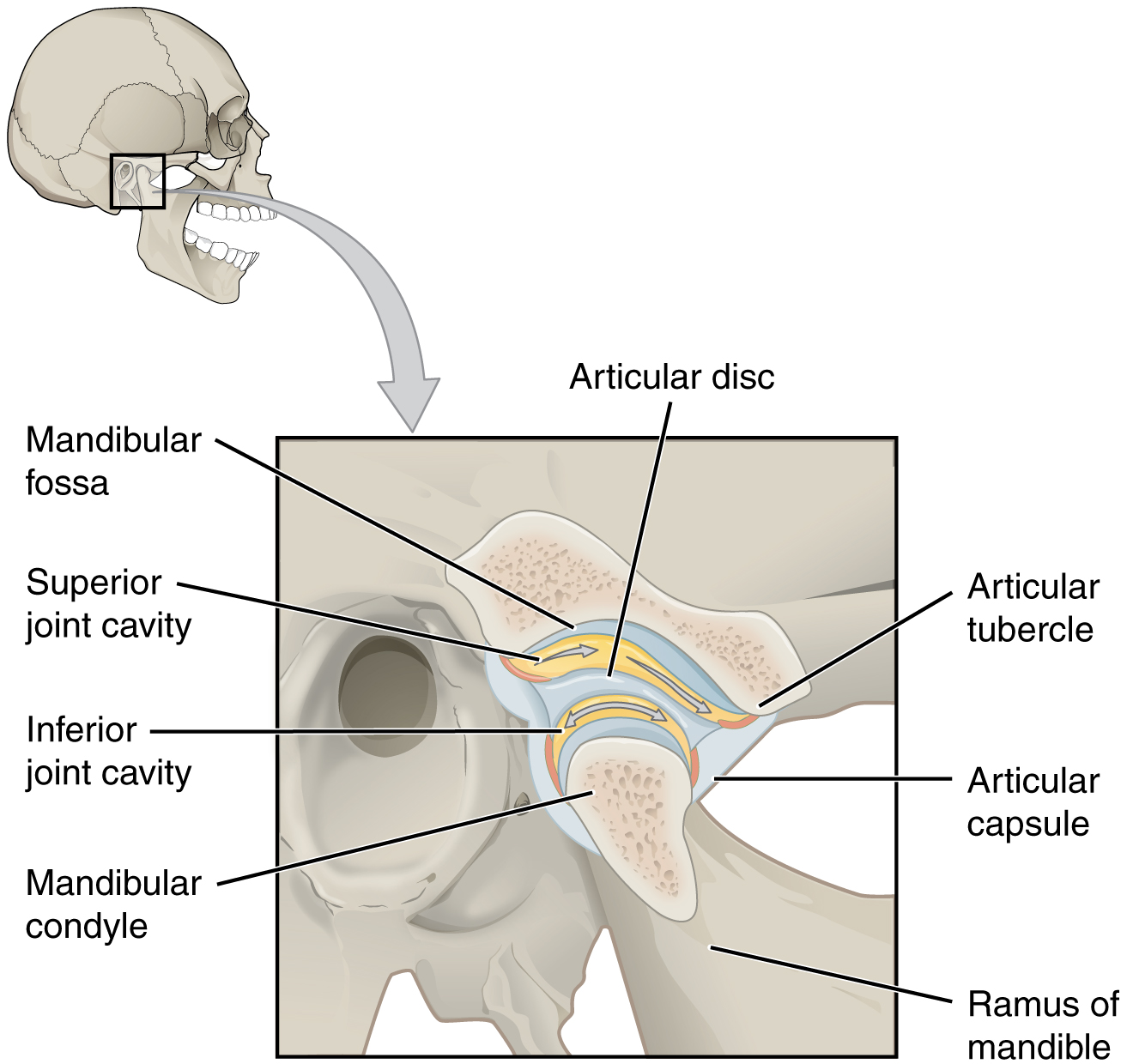
Identify the disc and the two joint compartments. Recall rotation versus translation, then connect the condyle and articular tubercle to dislocation and the disc to TMD assessment.
Temporomandibular joint · OpenStax College · CC BY 3.0. Original image; displayed at a responsive size. Original publication.
Why prostheses respond differently to contacts
| Restoration | Support behaviour | Occlusal concern |
|---|---|---|
| Tooth-supported fixed restoration | Teeth have periodontal mobility and sensory feedback | Material clearance, contact position and loading of weakened tissue. |
| Distal-extension RPD | Teeth and mucosa displace differently | Premature contacts can rotate the base and load terminal abutments. |
| Complete denture | The whole base can move on supporting mucosa | Tooth position and contact pattern influence tipping and stability. |
| Implant restoration | Support lacks a periodontal ligament | Loading, connector geometry and component stability require a deliberate plan. |
Neutral-zone record and putty index
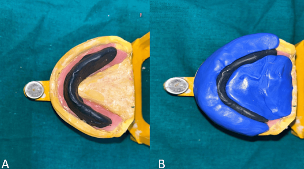
An additional functional recording technique used for the resorbed-ridge case. It guides tooth position and is not a compulsory stage for every complete denture.
Panel key
- A
- Neutral-zone record on the mandibular base.
- B
- Putty index made around the record.
Refining Complete Dentures — figure 5 · Rucha Chiddarwar, Anjali Bhoyar, Surekha A. Dubey, Mithilesh M. Dhamande and Sheetal R. Khubchandani · CC BY 4.0. Original published figure reproduced without alteration; panel letters and labels retained.
The four muscles of mastication
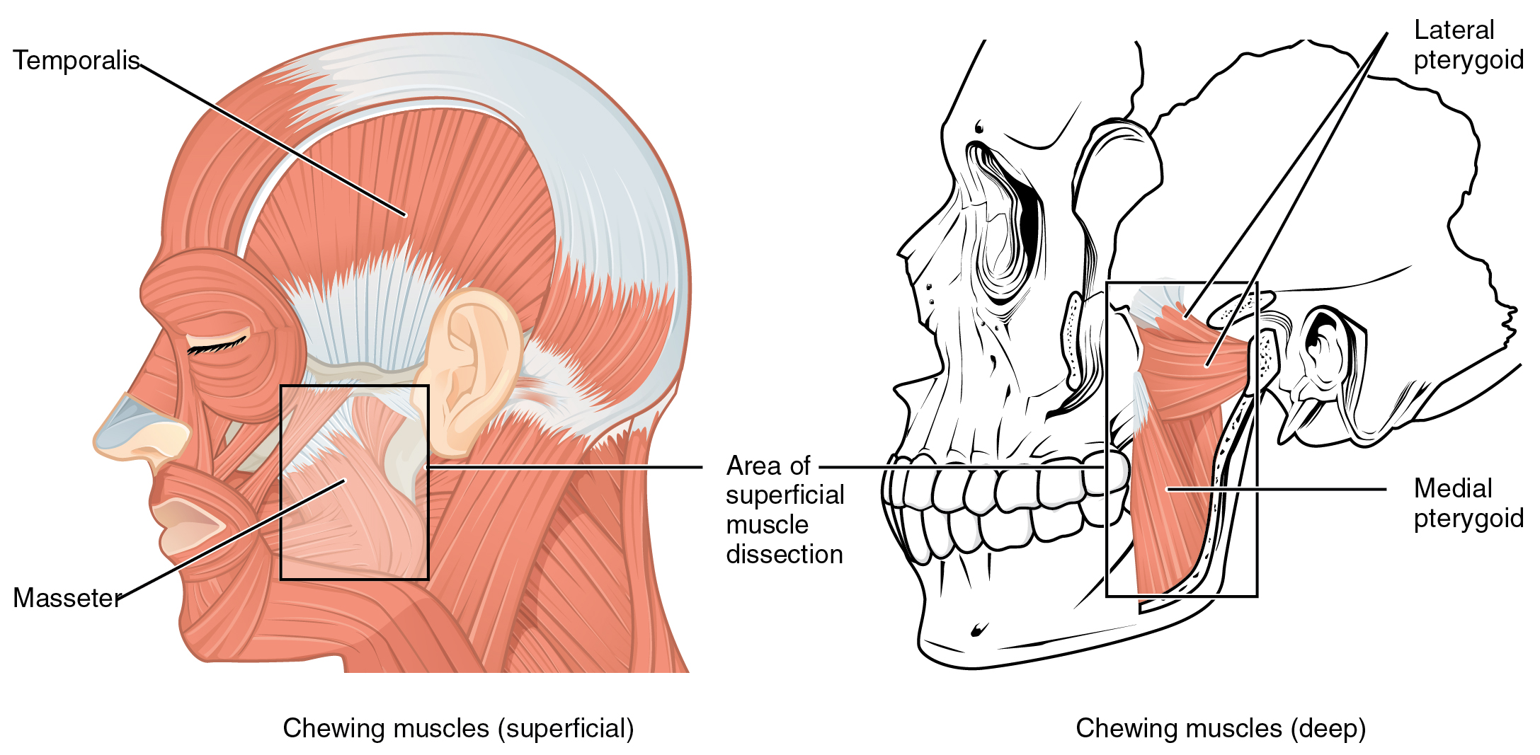
Identify temporalis and masseter in the superficial view, then both pterygoid muscles in the deeper view. Use the lines of pull to explain mandibular elevation, protrusion and side-to-side movement. All four receive motor supply from the mandibular division of the trigeminal nerve.
1108 Muscle that Move the Lower Jaw.jpg · OpenStax · CC BY 4.0. None; original image reproduced without alteration.
Which division of the trigeminal nerve supplies the muscles of mastication?
The mandibular division, V3. Do not confuse this with the facial nerve supply to the muscles of facial expression.
Intercuspal contacts and excursions
Assess contacts with the prosthesis fully seated. A denture rocking during examination can create a misleading mark and a false impression of tooth position. Examine movements as well as static closure, and ask whether a new contact destabilises the base or concentrates force on a cantilever or vulnerable restoration.
Canine guidance, group function and denture occlusal schemes describe different arrangements. Select a scheme in the clinical context rather than declaring one universal solution. Complete denture stability may justify a different approach from a single crown in an established natural dentition. The goal is a comfortable, reproducible and maintainable functional relationship.
Overjet and overbite
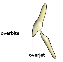
Overjet describes the horizontal relationship between the incisors; overbite describes their vertical overlap. Record the magnitude, direction and whether the overbite is complete or incomplete. The schematic is not drawn to a clinical measurement scale.
Overjet-overbite.png · Nielson2000 · CC BY-SA 3.0. None; original image reproduced without alteration.
Which measurement is horizontal: overjet or overbite?
Overjet is horizontal. Overbite is the vertical overlap of the incisors.
Vertical dimension, space and verification
Evaluate interarch space, tooth display, speech, facial support and available material thickness together. A small increase in vertical dimension may be part of selected rehabilitation, but must follow diagnosis and a planned verification strategy. Do not use a single rest-space measurement to justify an extensive change.
Diagnostic mounting, photographs, a wax-up or digital plan and trial restorations may help assess a complex proposed relationship. Check the clinical transfer: casts and virtual articulators are representations with limitations. Record the chosen reference position and how it was verified, and reassess after the restoration is functioning.
Worked problem: recurrent chipping
A ceramic bridge chips repeatedly near an excursion contact. Assess framework support, material thickness, connector design, surface damage, parafunction and actual contact position. The contact may contribute, but it is not the only possible explanation. Select an intervention that addresses the identified mechanism rather than repeatedly polishing the same site.
Where protection or occlusal adjustment is appropriate, explain its purpose and limits. Review the restoration and symptoms after treatment; removing every contact from a pontic or implant restoration without a coherent plan can compromise function.
Record a reference before reorganising
Centric relation and intercuspal position describe different reference concepts. The former is a jaw relationship independent of tooth contacts; the latter is the position of greatest tooth interdigitation. A patient can have a stable usable intercuspation without a need to reorganise every contact.
When a planned rehabilitation changes the reference or vertical relationship, document the reason and the verification method. Check the reproducibility of jaw records and stable support for the recording materials. An articulator or virtual representation can organise the plan, but cannot validate an unstable clinical record or predict every aspect of the patient’s adaptation.
A distinction to remember
Articulating-paper mark size is not a direct force measurement, and one occlusal scheme is not appropriate for every support system.
A complete lower denture rocks only when the patient closes. What would you examine?
Reveal the reasoning
Check contacts with both dentures fully seated, along with base fit, tooth position and the jaw record. A premature contact can tip the base. Correct the diagnosed cause rather than assuming every rocking denture needs a new impression or more adhesive.
Guidance to read alongside this chapter
Test your recall
How do intercuspal position and centric relation differ?
Show answer
Intercuspal position is determined by maximum tooth fit. Centric relation is a jaw relationship independent of tooth contact, used as a reference in appropriate planning. Identify which relationship is being recorded and why rather than treating the terms as interchangeable.