
Histopathology correlation
A tissue report answers a question about the specimen examined. Interpret it alongside the lesion, sampling method and clinical course, then make the result lead to an accountable action.
Quick revision summary
Describe the clinical question before sampling
Histopathology examines tissue architecture and cellular change. It can distinguish processes that overlap in appearance, but its answer depends on the tissue supplied and the clinical question.
Read the report’s scope and limitations
Identify the specimen, adequacy and diagnosis before interpreting terms such as dysplasia, invasion and margins. Epithelial dysplasia describes microscopic architectural and cellular abnormalities; it is not interchangeable with a clinical label such as leukoplakia.
Make result tracking part of the biopsy plan
Allocate responsibility for checking that the specimen arrives, obtaining the result, reviewing it and explaining the implications to the patient. Record the agreed follow-up before the patient leaves.
Common mistake
A reassuring report must be interpreted in relation to the exact tissue sampled and the remaining clinical findings.
Histopathology: connect the specimen, report and clinical findings
A tissue report answers a question about the specimen examined. Interpret it alongside the lesion, sampling method and clinical course, then make the result lead to an accountable action.
Describe the clinical question before sampling
Histopathology examines tissue architecture and cellular change. It can distinguish processes that overlap in appearance, but its answer depends on the tissue supplied and the clinical question. Record the exact site, duration, symptoms, distribution, changes over time and relevant exposures or medicines. A labelled photograph or site diagram can help the pathologist understand which part of a larger lesion was sampled.
An incisional biopsy samples part of a lesion; an excisional procedure aims to remove the lesion. These provide different information about extent and margins. A small sample showing inflammation does not prove that unsampled tissue is identical. Choose the sampling plan through the appropriate clinical pathway and competence, especially for suspected malignancy, a vascular lesion or a neck mass.
Oral mucosa: epithelium, basement membrane and lamina propria
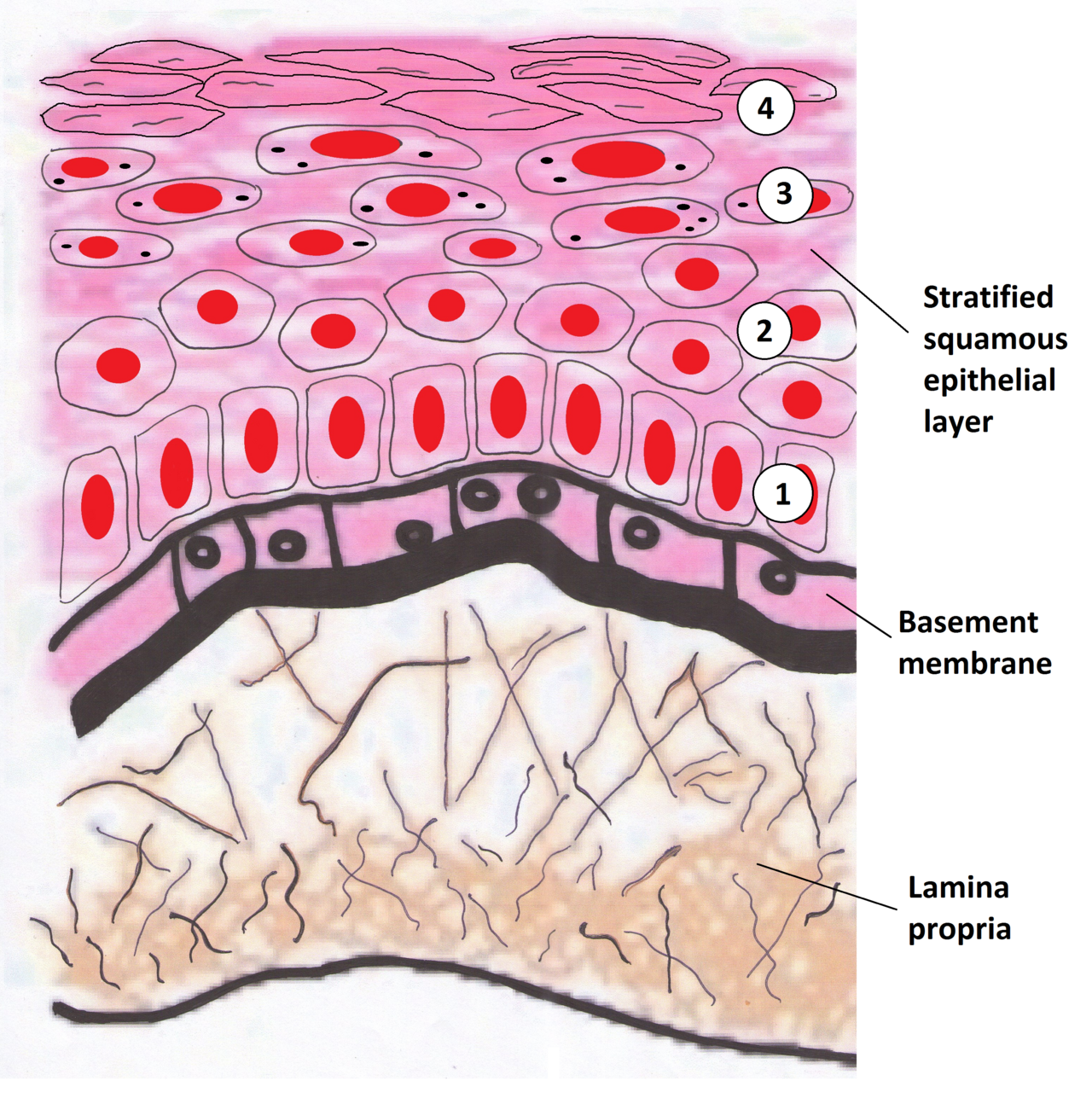
Identify the epithelium, basement membrane and lamina propria. Relate the epithelial–stromal interface to in-situ disease and invasion. This drawing represents a keratinising surface; oral sites have different keratinisation patterns. Use the printed anatomical names for revision.
Oral mucosa: epithelium, basement membrane and lamina propria · Wiki-minor (own work, 2011) · CC BY-SA 3.0. Unchanged published raster preview; displayed responsively. Original available from the source.
Select the correct specimen
| Question | Typical approach | Handling / caution |
|---|---|---|
| Routine mucosal histology | Representative incisional or appropriate excisional specimen | Formalin according to laboratory requirements; give site, history and differential. |
| Autoimmune blistering | Perilesional sample for direct immunofluorescence plus histology as advised | Arrange the transport medium; formalin alone is unsuitable for the DIF sample. |
| Bacterial / fungal culture | Suitable deep tissue or aspirate when indicated | Avoid a superficial contaminated swab as a substitute; separate microbiology handling. |
| Gland / neck mass | Imaging and specialist sampling strategy | Do not choose an open biopsy blindly; protect definitive management. |
| Suspected vascular lesion | Vascular assessment first | A casual incision may cause major bleeding. |
Leukoplakia: a focal white plaque
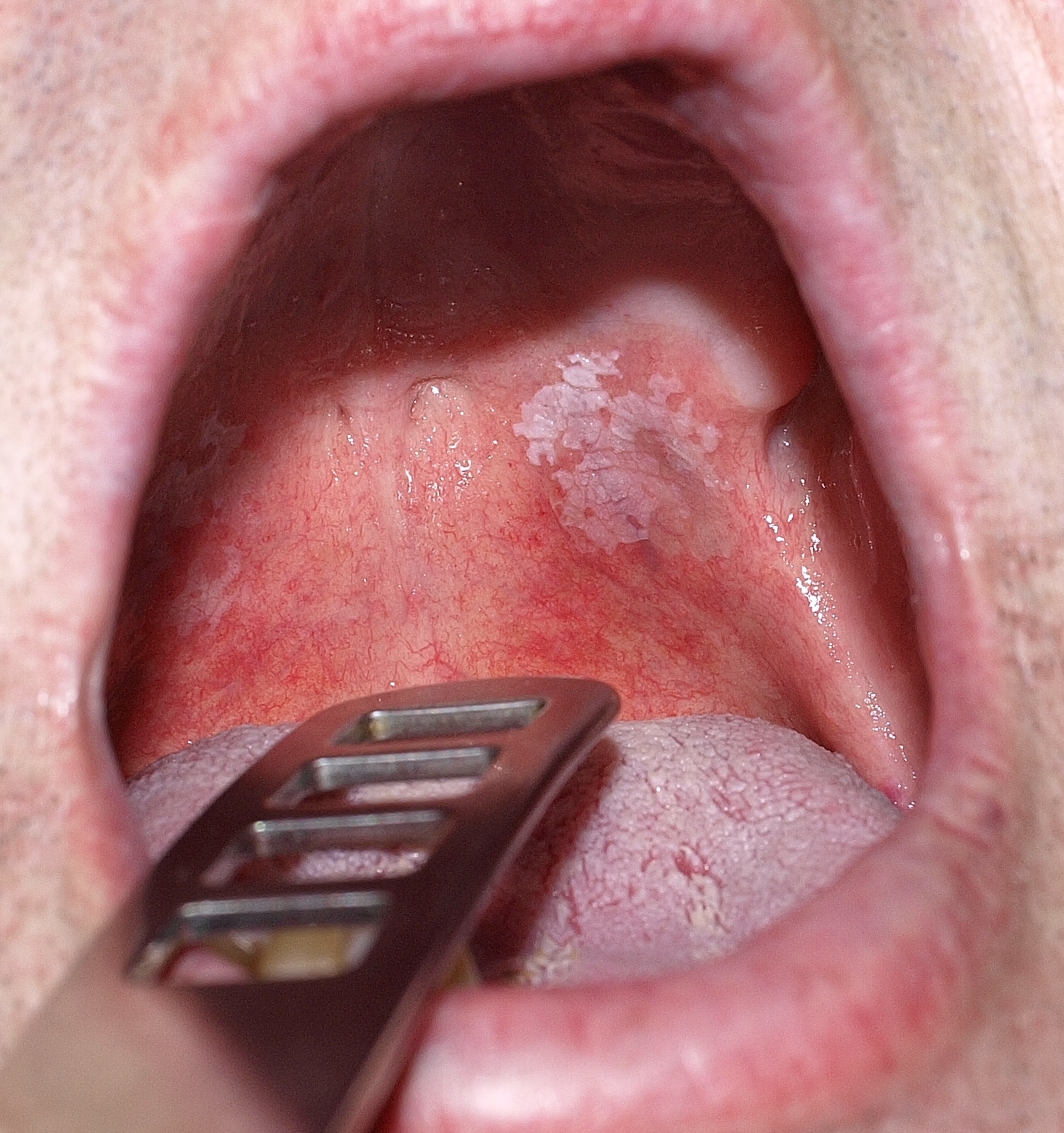
Describe the plaque’s site, outline, surface and surrounding mucosa before proposing a diagnosis. The source calls this lesion leukoplakia and reports that malignancy was excluded histologically in that case. That result cannot be inferred from its appearance or applied to another patient; white colour alone does not establish benignity.
Orale_Leukoplakie.jpg · Klaus D. Peter, Gummersbach, Germany · CC BY 3.0 DE. Source image reproduced without alteration; any arrows or circles are part of the original.
Can this photograph tell you whether the plaque contains epithelial dysplasia?
No. Dysplasia is a histological assessment. Clinical features guide the differential, urgency and sampling, but the photograph alone cannot grade or exclude it.
Protect the specimen’s diagnostic value
Representative viable tissue, adequate depth and minimal crush or thermal artefact improve the chance of a useful result. A necrotic ulcer centre alone may fail to show the underlying process. Heterogeneous lesions may need carefully selected sampling from more than one area. Label each site accurately and communicate orientation where relevant.
Check the laboratory’s requirements before taking an unusual specimen. Routine histology, direct immunofluorescence and microbiology require different handling. A specimen intended for immunofluorescence should not simply be put into formalin. The right clinical question with the wrong transport medium can still produce an unusable investigation.
Odontogenic keratocyst: a posterior mandibular example
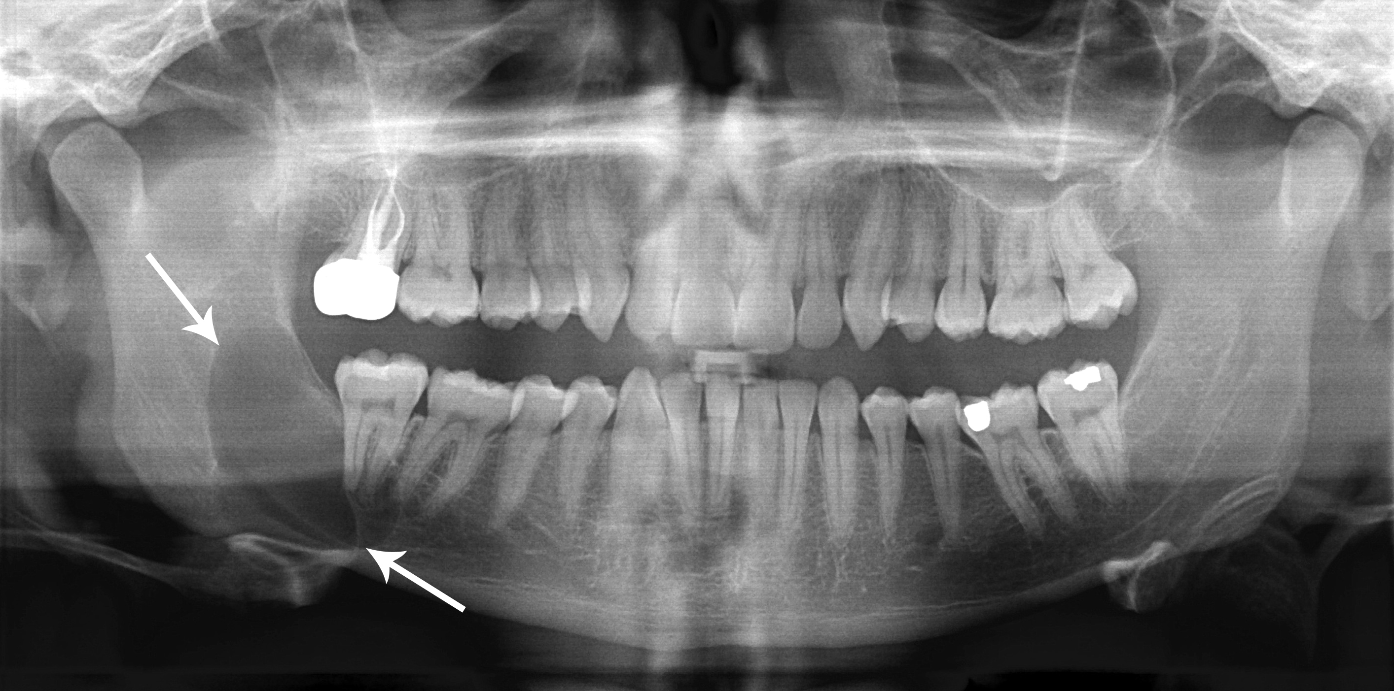
Use the original arrows to locate the posterior mandibular radiolucency and inspect its extent. The source diagnosis is an odontogenic keratocyst, described under the historical name “keratocystic odontogenic tumour”. Its relatively well-defined appearance does not make follow-up unnecessary. Other jaw lesions can look similar; definitive classification requires correlation with tissue findings.
Classic_keratocystic_odontogenic_tumour.jpg · Coronation Dental Specialty Group · CC BY 3.0. Source image reproduced without alteration; any arrows or circles are part of the original.
Can the relatively well-defined border distinguish this keratocyst from every other odontogenic cyst?
No. Borders and location help rank possibilities but overlap between lesions. The source diagnosis is supplied for teaching; it cannot be independently established from this panoramic image alone.
Read the report’s scope and limitations
Identify the specimen, adequacy and diagnosis before interpreting terms such as dysplasia, invasion and margins. Epithelial dysplasia describes microscopic architectural and cellular abnormalities; it is not interchangeable with a clinical label such as leukoplakia. Invasion has a different meaning and changes the treatment pathway. A report applies to the tissue examined, not automatically to the whole lesion.
Check whether the result answers the original question. “No malignancy identified” in a superficial sample is different from confident exclusion in an adequate, representative investigation. When the report does not explain persistent induration, ulceration or progression, discuss the discrepancy with the pathologist and appropriate specialist. Further assessment or sampling may be required.
Fibrous dysplasia, osteoma and cementoblastoma
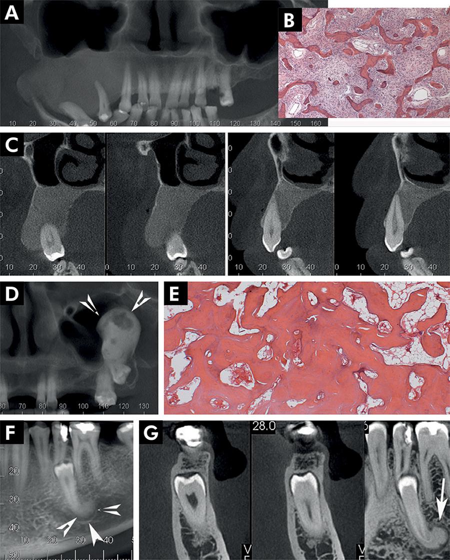
Compare the margins and root relationships of these source cases.
Panel key
- A–C
- Fibrous dysplasia.
- D–E
- Central osteoma.
- F–G
- Cementoblastoma.
Periapical radiopaque-hyperdense jaw lesions 3.jpg · Brunno Santos Freitas Silva, Mike Reis Bueno, Fernanda P. Yamamoto-Silva, Ricardo Santiago Gomez, Ove Andreas Peters and Carlos Estrela · CC BY 4.0. Reproduced without alteration. Labels, arrows and circles, where present, belong to the source image. Original publication.
Which relationship deserves attention in the cementoblastoma panels?
The lesion’s attachment to the root.
Make result tracking part of the biopsy plan
Allocate responsibility for checking that the specimen arrives, obtaining the result, reviewing it and explaining the implications to the patient. Record the agreed follow-up before the patient leaves. A result that is filed without interpretation has not completed the investigation.
Communicate significant findings with the urgency they require and ensure referral or further treatment is arranged. If the patient misses a result appointment, follow the appropriate contact and escalation process. Explain uncertainty in understandable terms: the sample may be nondiagnostic, may represent only one part of a lesion or may need further specialist review. Avoid presenting a provisional or limited report as a final guarantee.
Ameloblastoma: epithelial islands on histology
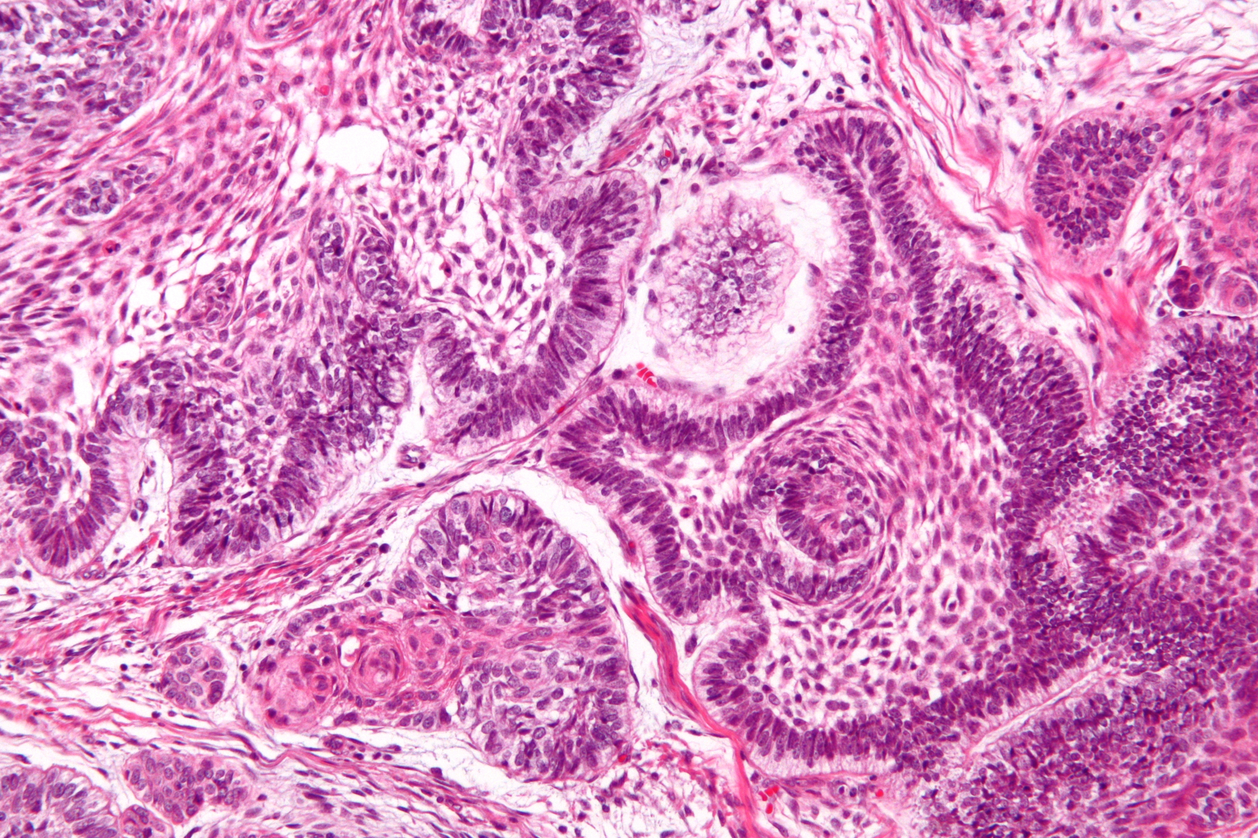
Locate the epithelial islands within the connective-tissue stroma. The source identifies peripheral palisading, reverse nuclear polarisation and central stellate-reticulum-like cells. This is a separate specimen from the CT teaching case. A complete diagnosis considers the sampled architecture and the clinical and radiographic findings.
Ameloblastoma - high mag.jpg · Nephron · CC BY-SA 3.0. Reproduced without alteration. Labels, arrows and circles, where present, belong to the source image.
What does reverse nuclear polarisation refer to in this teaching example?
The nuclei of peripheral cells lie away from the basement-membrane side. It is interpreted with the wider architecture rather than treated as an isolated diagnostic rule.
Odontoma: dental hard-tissue architecture
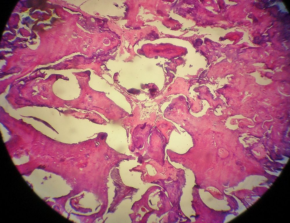
The source identifies an odontoma. Inspect how the dental tissues are organised and compare this tissue view with the atlas’s radiographic odontoma panels, which show different teaching cases. Imaging describes the mineralised pattern and tooth relationships; histology assesses the tissue architecture.
Odontoma.jpg · JXtra · CC BY-SA 3.0. Reproduced without alteration. Labels, arrows and circles, where present, belong to the source image.
Why keep the radiographic and histological descriptions distinct?
They describe different aspects of the lesion. A radiopaque pattern is not itself a microscopic diagnosis, and the specimen must be interpreted with the case information.
Mucocele: low-power tissue appearance
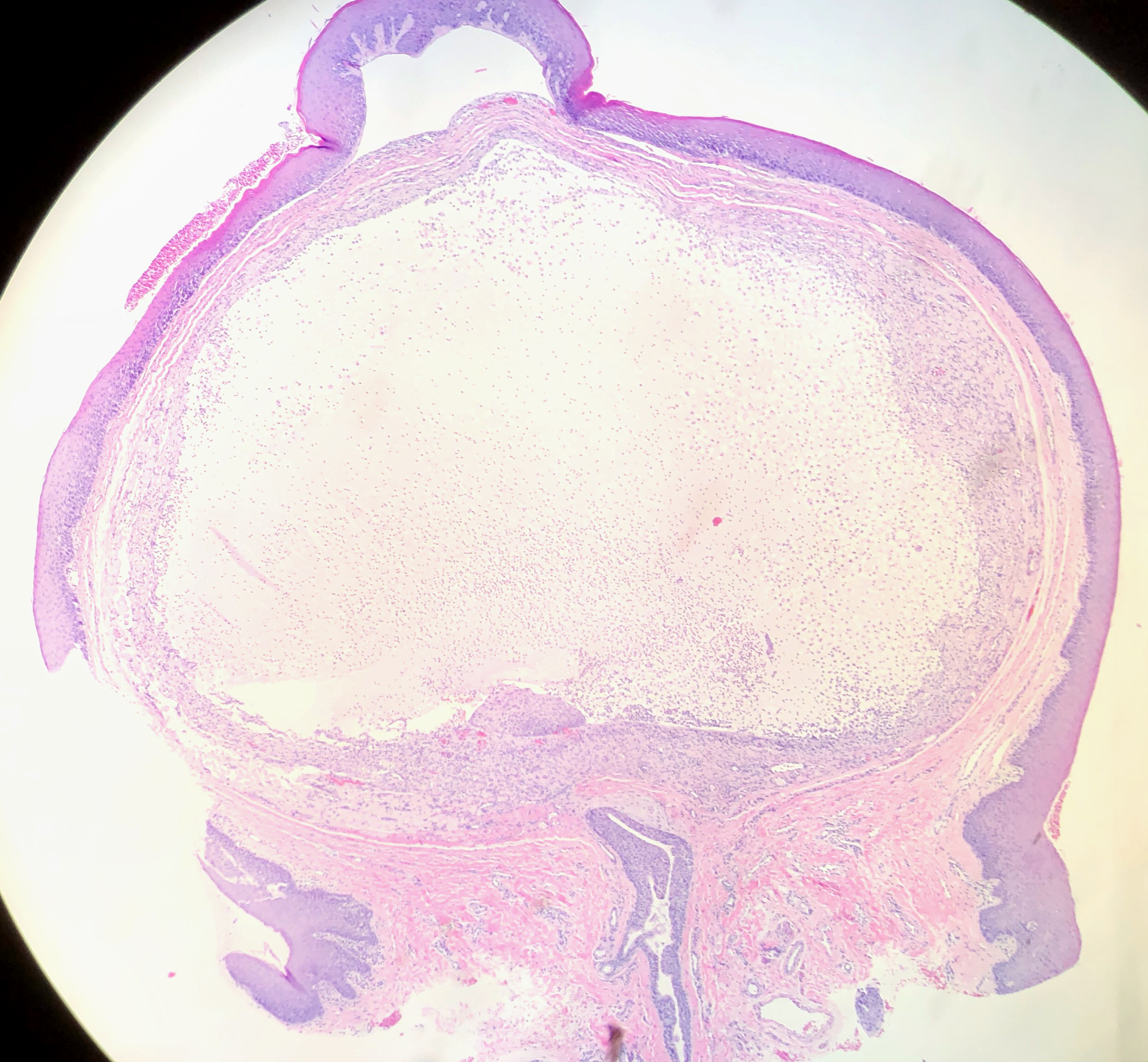
The source identifies a lower-lip mucocele. Orient yourself at the surface epithelium, then find the large pale mucin-rich area beneath it and the surrounding tissue reaction. Distinguishing mucous extravasation from a duct-lined retention lesion requires attention to the complete specimen and report.
Mucocele of Lower Lip (49425670122).jpg · Ed Uthman · CC BY 2.0. Reproduced without alteration. Labels, arrows and circles, where present, belong to the source image.
Which tissue relationship is important when interpreting a mucocele specimen?
The relationship of mucin, the surrounding reaction and any duct or epithelial lining. A cavity-like space alone does not establish that it is a true epithelial-lined cyst.
Radicular cyst: epithelial metaplasia
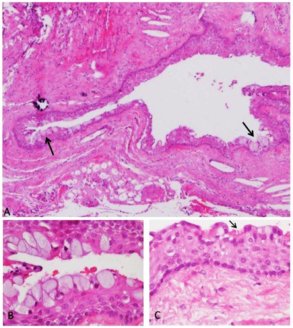
The source demonstrates metaplastic epithelial changes in a radicular-cyst wall. These are selected examples, not a description of every radicular cyst. Interpret unusual lining features with the rest of the architecture and the clinical relationship to the teeth.
Panel key
- A
- Cyst wall with mucous-cell changes.
- B
- Higher-power view of mucous cells.
- C
- Ciliated epithelial cells, marked by the source arrow.
Histopathology of a periapical cyst, with metaplastic changes of mucous secreting cells, and ciliated cells.jpg · Igor Tsesis, Eyal Rosen, Liz Dubinsky, Amos Buchner and Marilena Vered · CC BY 2.5. Reproduced without alteration. Labels, arrows and circles, where present, belong to the source image. Original publication.
Do mucous cells alone establish a glandular odontogenic cyst?
No. Mucous-cell metaplasia can occur in other cysts. Classification requires the complete histological pattern and clinical correlation.
A distinction to remember
A reassuring report must be interpreted in relation to the exact tissue sampled and the remaining clinical findings.
A suspicious ulcer remains indurated after a biopsy reports nonspecific inflammation. What should happen?
Reveal the reasoning
Review the clinical–pathological fit and specimen adequacy, discuss with the appropriate specialist / pathologist and arrange further assessment or representative re-sampling. Do not discharge solely on a nonspecific report.
Guidance to read alongside this chapter
Test your recall
Can a small superficial biopsy exclude deeper disease?
Show answer
Not reliably if the sampled tissue is unrepresentative.