
Oral ulceration
Oral ulceration is loss of surface epithelium, with many possible causes. The useful starting distinction is a recurrent episode that heals completely versus a single persistent lesion. Combine pattern, site, duration and systemic findings, while protecting nutrition and avoiding delayed investigation of a suspicious abnormality.
Quick revision summary
Describe what is present
Record number, exact site, dimensions, shape, base, border and surrounding tissue. Palpate for induration or fixation and assess relevant lymph nodes. Document pain and effect on eating, drinking, speech and oral hygiene.
Ask about the rest of the patient
Relevant features include gastrointestinal symptoms, weight loss, genital ulceration, eye inflammation, skin lesions, recurrent infections and medicine changes. Behçet’s disease, inflammatory bowel disease, coeliac disease, neutropenia and immune compromise are among the context-dependent possibilities.
Review healing and explain the safety net
Arrange a defined reassessment when treating a presumed traumatic or inflammatory ulcer. Record whether the original lesion healed fully, decreased, persisted or changed. Improvement in pain alone does not prove that an abnormal area has resolved; inspect and palpate it again.
Common mistake
A history of recurrent ulcers does not explain a new persistent indurated lesion. Three weeks is a referral trigger, not a compulsory waiting period.
Oral ulceration: recognise patterns and investigate persistence
Oral ulceration is loss of surface epithelium, with many possible causes. The useful starting distinction is a recurrent episode that heals completely versus a single persistent lesion. Combine pattern, site, duration and systemic findings, while protecting nutrition and avoiding delayed investigation of a suspicious abnormality.
Describe what is present
Record number, exact site, dimensions, shape, base, border and surrounding tissue. Palpate for induration or fixation and assess relevant lymph nodes. Document pain and effect on eating, drinking, speech and oral hygiene. A diagram or photograph establishes a baseline and helps distinguish recurrence at a new site from one ulcer that never healed.
Ask about onset, preceding vesicles, trauma, previous episodes, complete healing, medicines and systemic illness. “Mouth ulcer” is a symptom description; a patient with a history of aphthae can still develop cancer, infection or a drug-related lesion.
Oral lichen planus: a mixed red and white appearance
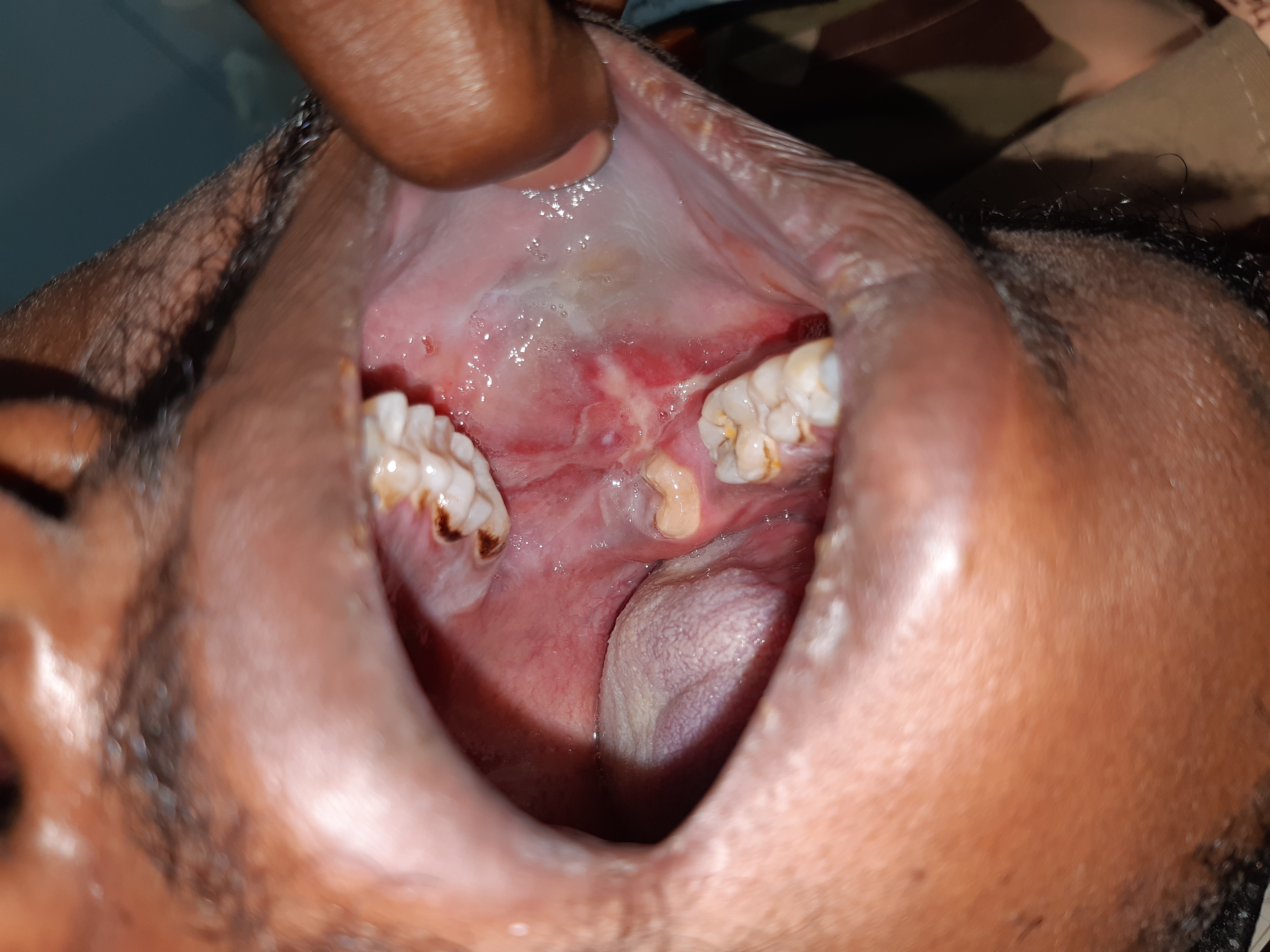
Compare the white striae and thicker white areas with the adjacent erythematous mucosa. The source labels this case oral lichen planus. A red or ulcerated area needs assessment in its own right: symptoms, change, induration and a persistent focal abnormality matter, even when a background lichenoid pattern is familiar.
Lichen_planus-intra_oral_lesion.jpg · Syed Wali Peeran · CC BY-SA 4.0. Source image reproduced without alteration; any arrows or circles are part of the original.
Does recognising a lichenoid pattern explain every red area in the photograph?
No. Describe and assess each significant area. Lichenoid disease, trauma, infection and dysplasia can overlap clinically; a persistent or changing focal lesion may need specialist assessment and biopsy.
Compare aphthous patterns
Recurrent aphthous stomatitis typically affects non-keratinised mucosa and has ulcer-free intervals. Minor lesions are usually small and heal without scarring; major ulcers are larger, deeper and more persistent and may scar. Herpetiform aphthae are numerous tiny ulcers that can merge, despite having no causal relationship to herpes virus.
Aphthous-like ulceration can accompany iron, folate or vitamin B12 deficiency and systemic disorders. Severe, new adult-onset, unusually persistent or function-limiting disease deserves directed investigation. Do not assume that every recurrent ulcer indicates a nutritional deficiency or needs a broad untargeted test panel.
Aphthous ulcer: labial mucosa
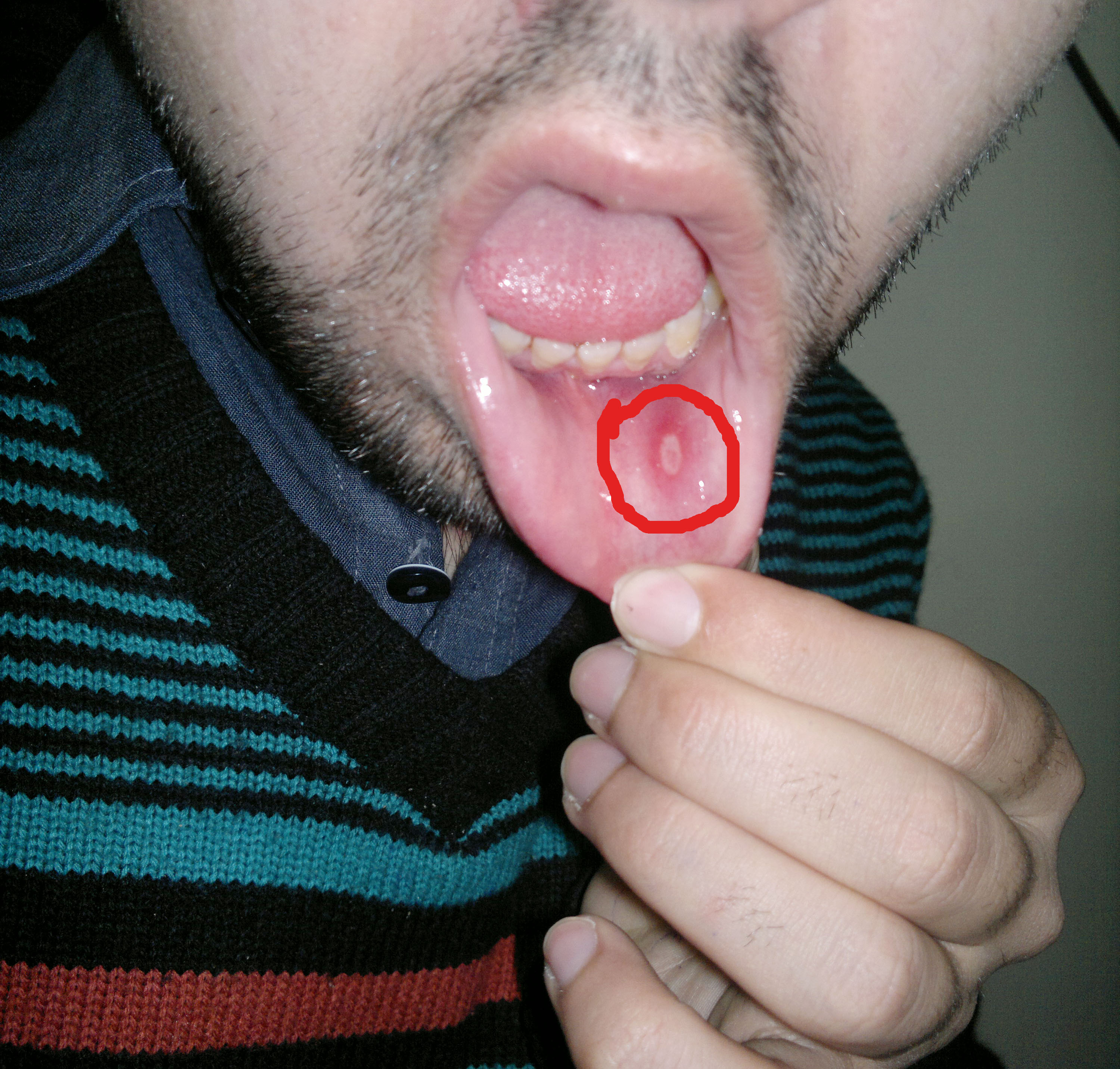
Locate the oval ulcer within the original red circle. Its pale floor and erythematous surround illustrate an aphthous appearance on labial mucosa. The history of recurrence, duration, healing and symptoms elsewhere helps distinguish recurrent aphthous stomatitis from other causes of ulceration.
Aphthous stomatitis on the labial mucosa.jpg · Farhan 9909 · CC BY-SA 4.0. Reproduced without alteration. Labels, arrows and circles, where present, belong to the source image.
What information would make an aphthous explanation more convincing?
A compatible history of recurrent, self-healing episodes and distribution, interpreted with examination. A persistent unexplained ulcer still needs assessment even if it resembles an earlier aphthous lesion.
Separate trauma, infection and immune disease
Mechanical trauma from a sharp tooth, denture or appliance should have a convincing relationship to the site. Chemical and thermal injury can also ulcerate mucosa. Remove a plausible cause and confirm healing; an incidental sharp cusp must not become an explanation for indefinite persistence.
Primary herpetic gingivostomatitis can cause widespread painful gingival and mucosal disease with systemic symptoms. Recurrent intraoral HSV often has a different distribution, commonly keratinised mucosa in an immunocompetent person. Immune status can change patterns. Widespread erosions or desquamative gingivitis may reflect lichen planus, pemphigus or pemphigoid rather than isolated ulcers.
Lower-lip squamous cell carcinoma
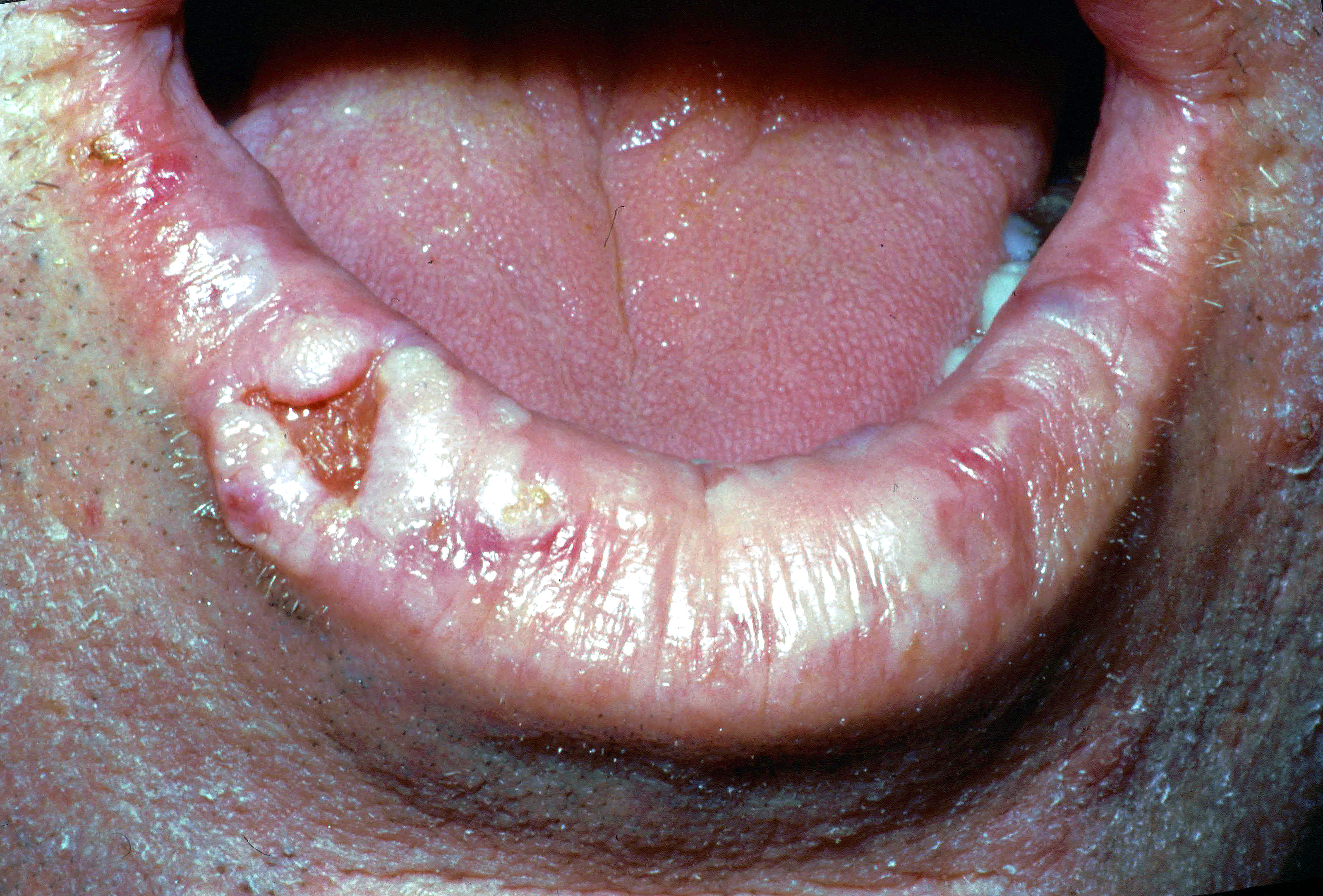
The source diagnosis is squamous cell carcinoma of the right lower lip. Describe the irregular ulcerated area, raised tissue and surrounding lip change. A clinical assessment also needs palpation, duration, progression, nodes and relevant exposures. A photograph cannot establish tumour depth or stage.
Lower lip cancer.jpg · Coronation Dental Specialty Group · CC BY-SA 4.0. Reproduced without alteration. Labels, arrows and circles, where present, belong to the source image.
Which important examination findings cannot be judged reliably from this photograph?
Induration, fixation, depth and regional lymph-node findings require clinical assessment. Tissue diagnosis and appropriate staging investigations add information that a surface photograph cannot supply.
Ask about the rest of the patient
Relevant features include gastrointestinal symptoms, weight loss, genital ulceration, eye inflammation, skin lesions, recurrent infections and medicine changes. Behçet’s disease, inflammatory bowel disease, coeliac disease, neutropenia and immune compromise are among the context-dependent possibilities. Oral findings do not establish any one systemic diagnosis.
Assess hydration, nutrition and general condition, especially in children, older people or immunosuppression. Inability to drink, systemic deterioration or extensive disease can require urgent medical care. Eye symptoms with mucocutaneous disease may need prompt ophthalmic assessment and should not be managed solely with a mouthwash.
Choose investigations and referral deliberately
For recurrent or atypical disease, selected blood tests can include a full blood count and haematinics, with coeliac or other testing guided by the history. Consider microbiology when infection is a specific question. A biopsy may be needed for a persistent focal ulcer or an uncertain inflammatory pattern.
NICE’s unexplained oral-ulcer criterion is persistence beyond three weeks; suspicious findings can justify referral earlier. Use the appropriate local suspected-cancer pathway, not repeated empirical treatment. This threshold is a trigger for action, not a reason to wait when induration, progressive change or other red flags already create concern.
Treat symptoms without losing the diagnosis
Remove confirmed local trauma, provide suitable analgesia and support gentle hygiene and hydration. Topical corticosteroids can help appropriately diagnosed inflammatory or aphthous disease, selected and reviewed through the current prescribing pathway. They do not establish the cause and may worsen an untreated infection.
Antivirals, antifungals or systemic immune therapy have specific indications and are not interchangeable ulcer treatments. Avoid routine antibiotics for uncomplicated aphthae or trauma. Check medicine interactions, age restrictions and the patient’s medical context before prescribing. Severe disease may need specialist care rather than stronger unsupervised symptomatic treatment.
Review healing and explain the safety net
Arrange a defined reassessment when treating a presumed traumatic or inflammatory ulcer. Record whether the original lesion healed fully, decreased, persisted or changed. Improvement in pain alone does not prove that an abnormal area has resolved; inspect and palpate it again.
Explain the likely course and what requires earlier contact, including inability to maintain fluids, systemic illness, spreading lesions, eye problems or a new lump. Persistent or changing disease should reopen the differential. The follow-up plan should name responsibility for test results, specialist referral and communication to the patient.
A distinction to remember
A history of recurrent ulcers does not explain a new persistent indurated lesion. Three weeks is a referral trigger, not a compulsory waiting period.
A lateral tongue ulcer has persisted for five weeks beside a sharp tooth. What should you do?
Reveal the reasoning
Assess and address the local trauma, but do not let it delay appropriate suspected-cancer referral for an unexplained persistent ulcer, especially with suspicious findings. Record size, induration and nodes. A nearby sharp tooth does not prove the cause of a non-healing lesion.
Test your recall
Which history feature most clearly separates recurrence from persistence?
Show answer
Establish whether each ulcer heals completely and whether there are genuinely ulcer-free intervals. Several episodes at different sites suggest a recurrent process; one lesion that never heals needs a different assessment. Patients may use “keeps coming back” for either pattern.