
Pulpal and apical diagnosis
A single test result should not become the whole diagnosis.
THE ANATOMY LAB / INTERACTIVE 3DStudy the pulp space with a guided 3D view↗Quick revision summary
Interpret pulp and apical findings together
Reversible pulpitis typically produces stimulus-related symptoms that settle; symptomatic irreversible pulpitis may cause lingering thermal or spontaneous pain. Clinical categories are imperfect descriptions of tissue state and should be used with the full case.
Understand what each test measures
Cold and electric tests assess a neural response, so they are sensibility tests rather than direct measurements of blood supply. Compare with control teeth and consider recent trauma, immature roots, calcification and restorations when a result is absent or uncertain.
Resolve competing sources before intervention
Consider cracks, periodontal disease, occlusal trauma and non-odontogenic pain when the findings do not fit. A narrow isolated probing defect, pain on release of biting pressure or symptoms after restoration can direct further assessment, but none is a stand-alone diagnosis.
Common mistake
A familiar radiographic pattern cannot replace separate, evidence-based pulpal and apical assessments.
Pulpal and apical diagnosis as two linked decisions
A single test result should not become the whole diagnosis.
Interpret pulp and apical findings together
Reversible pulpitis typically produces stimulus-related symptoms that settle; symptomatic irreversible pulpitis may cause lingering thermal or spontaneous pain. Clinical categories are imperfect descriptions of tissue state and should be used with the full case. Necrosis is supported by the pattern of tests and findings, not one absent response. Trauma, immature roots, calcification and restorations can change test reliability.
Apical diagnosis considers tenderness to percussion or palpation, swelling, sinus tracts and imaging. Symptomatic apical periodontitis can occur without a visible radiolucency. A chronic apical abscess may drain and be relatively painless. Separate the patient’s symptom pattern from the tissue diagnosis, and distinguish an endodontic source from periodontal, cracked-tooth and non-dental causes.
Apical radiolucency around a premolar
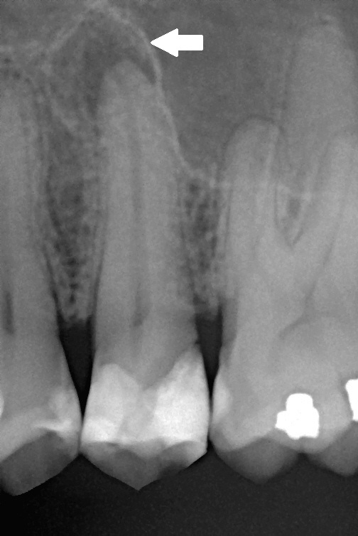
The original arrow marks the radiolucency described as apical periodontitis in the source. Relate this finding to symptoms and pulp tests rather than assigning a diagnosis from the shadow alone.
Chronic apical periodontitis.jpg · Michele Gardini; original arrow annotation by Lesion · CC BY-SA 3.0. Image reproduced without alteration; original marks and any embedded credits retained.
Tooth & supporting tissues
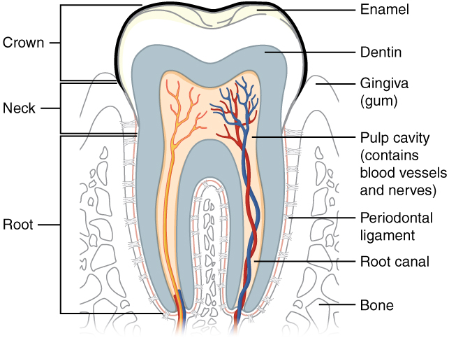
Trace the pulp and root canal to the apical region, then identify the periodontal ligament and supporting bone. Relate these structures to avulsion, apical pathology and extraction. The original uses the spelling “dentin”; cementum is not separately labelled.
Tooth & supporting tissues · OpenStax College · CC BY 3.0. Original image; displayed at a responsive size. Original publication.
Key distinctions in practice
| Finding or option | What it means | Clinical consequence |
|---|---|---|
| Cold / electrical test | Sensibility rather than direct perfusion | Compare control teeth and repeat equivocal results. |
| Percussion | Apical supporting-tissue response | Does not alone establish pulp vitality. |
| Probing | Periodontal pattern | An isolated deep defect may suggest a fracture or local lesion. |
| Radiography | Anatomy and mineralised-tissue change | Early disease can be radiographically occult. |
The pulp–dentine interface
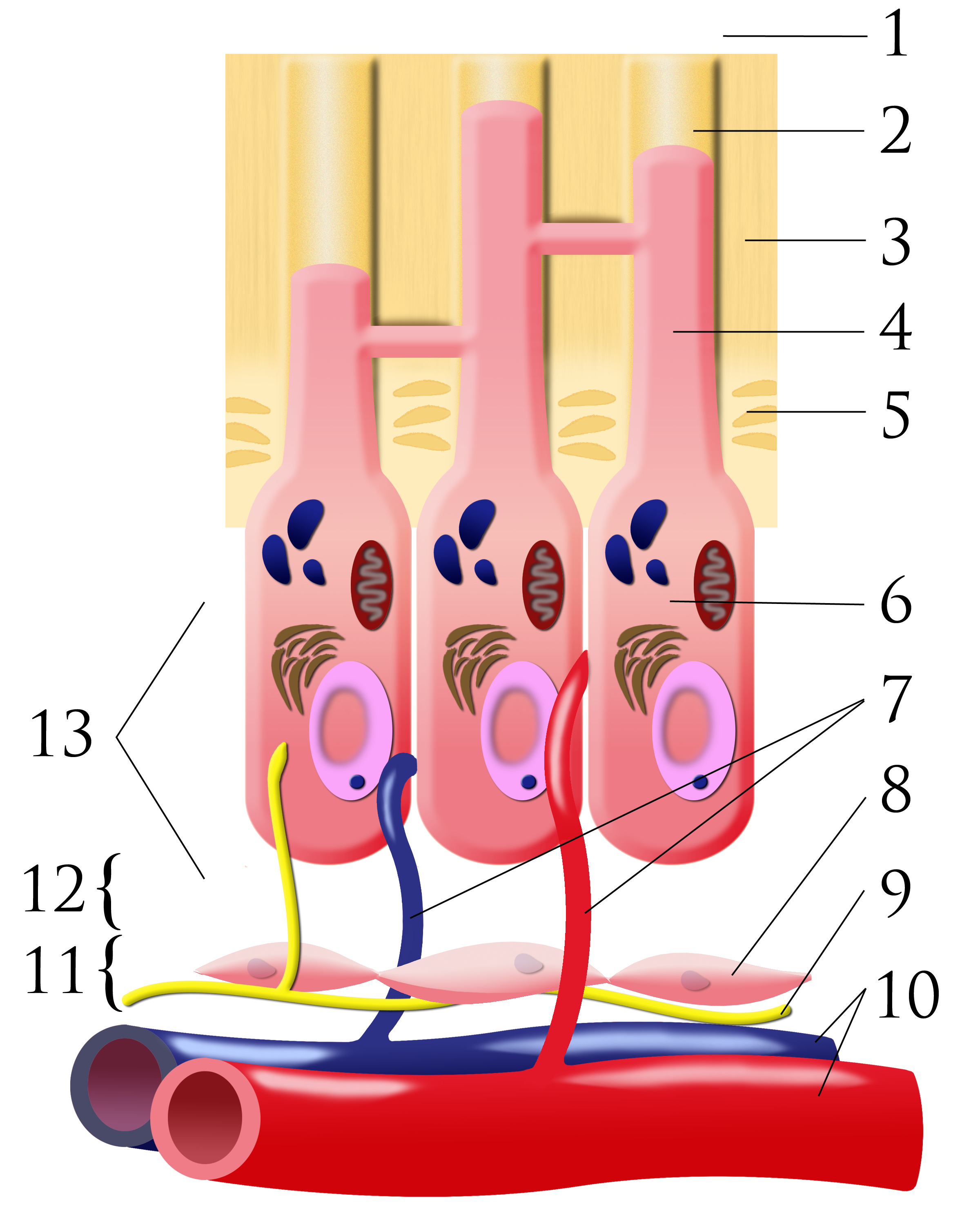
Number key: 1 outer-tooth direction; 2 dentinal tubule; 3 dentine; 4 odontoblastic process; 5 predentine; 6 odontoblast; 7 capillaries; 8 fibroblasts; 9 nerve; 10 artery and vein; 11 cell-rich zone; 12 cell-poor zone; 13 pulp chamber. This is a conceptual schematic, not a scale histological section.
Pulpal dentin junction.png · Ian Furst · CC BY-SA 3.0. None; original image reproduced without alteration.
Which cells form dentine, and where are their cell bodies?
Odontoblasts form dentine. Their cell bodies line the pulp–dentine border, with processes extending into dentinal tubules.
Understand what each test measures
Cold and electric tests assess a neural response, so they are sensibility tests rather than direct measurements of blood supply. Compare with control teeth and consider recent trauma, immature roots, calcification and restorations when a result is absent or uncertain. A positive response does not describe the histological extent of inflammation, and one negative response does not automatically establish necrosis.
Percussion and palpation assess the response of supporting tissues. A tooth can be tender to percussion while the pulp still responds. Conversely, a necrotic pulp may be relatively symptomless. Record the test, comparison and response rather than a bare tick or a diagnostic label unsupported by the findings. Different pulpal and apical conclusions should be documented separately.
Periapical sclerosis: compare four patterns
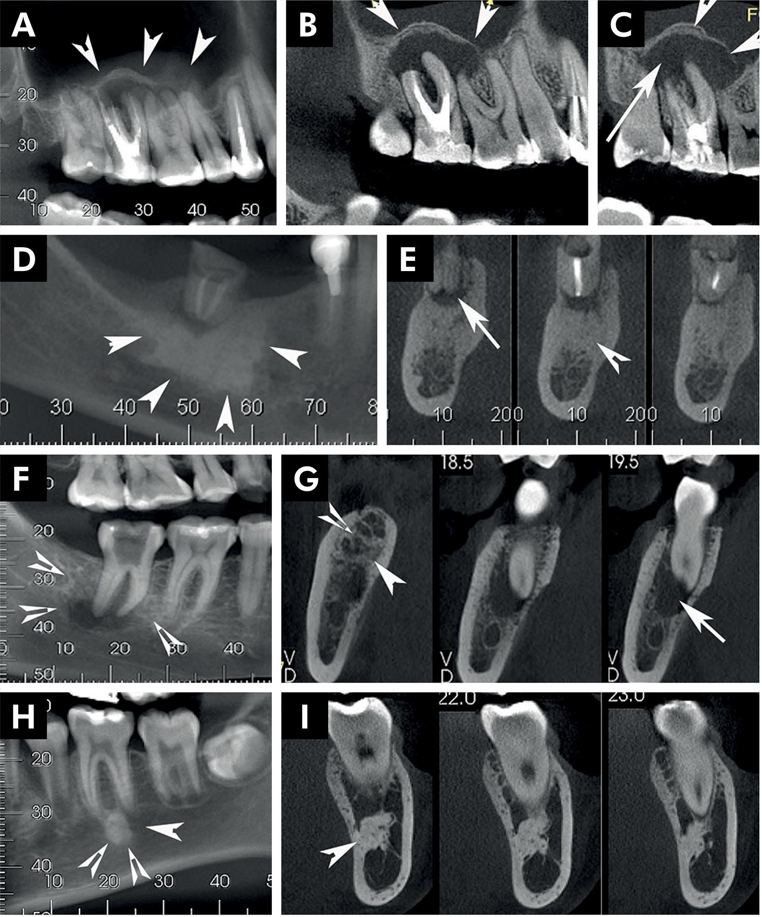
Compare the source diagnoses below. Tooth findings and lesion distribution help interpret the opacity.
Panel key
- A–C
- Reactive bone associated with inflammatory change.
- D–E
- Condensing osteitis.
- F–G
- Chronic osteomyelitis.
- H–I
- Idiopathic osteosclerosis.
Periapical radiopaque-hyperdense jaw lesions 1.jpg · Brunno Santos Freitas Silva, Mike Reis Bueno, Fernanda P. Yamamoto-Silva, Ricardo Santiago Gomez, Ove Andreas Peters and Carlos Estrela · CC BY 4.0. Reproduced without alteration. Labels, arrows and circles, where present, belong to the source image. Original publication.
Does periapical opacity alone establish an endodontic diagnosis?
No. Correlate it with tooth assessment and the broader pattern.
Resolve competing sources before intervention
Consider cracks, periodontal disease, occlusal trauma and non-odontogenic pain when the findings do not fit. A narrow isolated probing defect, pain on release of biting pressure or symptoms after restoration can direct further assessment, but none is a stand-alone diagnosis. Review restorability as well as the source of pain.
Radiographs show mineralised structures and projected anatomy; early apical inflammation may have no visible change. A radiolucency near an apex may represent anatomy or non-endodontic disease, especially when the clinical findings disagree. Use justified additional views or specialist advice when needed. Explain uncertainty and arrange reassessment rather than treating an image as sufficient justification for root canal treatment.
Cemento-osseous dysplasia: localised and florid examples
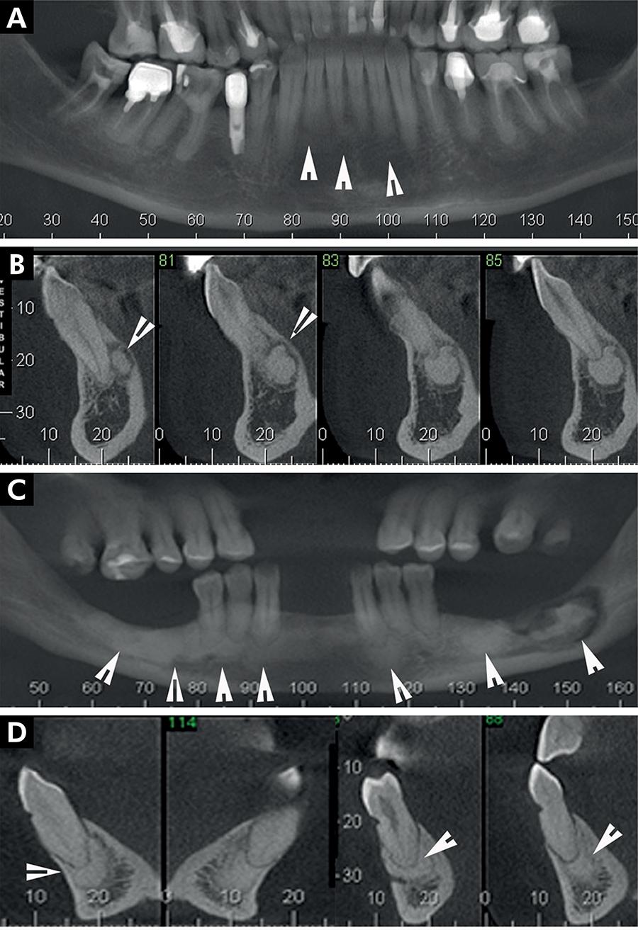
Compare the distribution and separation from roots in the source examples.
Panel key
- A–B
- Periapical cemento-osseous dysplasia.
- C–D
- Florid cemento-osseous dysplasia; the source also describes sequestration in C.
Periapical radiopaque-hyperdense jaw lesions 2.jpg · Brunno Santos Freitas Silva, Mike Reis Bueno, Fernanda P. Yamamoto-Silva, Ricardo Santiago Gomez, Ove Andreas Peters and Carlos Estrela · CC BY 4.0. Reproduced without alteration. Labels, arrows and circles, where present, belong to the source image. Original publication.
Why assess the teeth before attributing these findings to pulp disease?
The imaging distribution may have a non-endodontic explanation.
A distinction to remember
A familiar radiographic pattern cannot replace separate, evidence-based pulpal and apical assessments.
A tooth has lingering cold pain and percussion tenderness but no apical radiolucency. What is the lesson?
Reveal the reasoning
Pulpal and apical symptoms can precede visible bone changes. A normal film does not exclude either symptomatic pulpitis or symptomatic apical periodontitis.
Guidance to read alongside this chapter
Test your recall
Can a periapical radiolucency alone establish pulp status?
Show answer
No; it must be interpreted with clinical tests and history.