
Simple extraction
An apparently straightforward extraction still requires a diagnosis, a justified indication and a plan for unexpected difficulty. Understand the periodontal ligament, root shape and surrounding bone before deciding how the tooth can move. The endpoint is safe treatment with controlled bleeding and an informed recovery plan.
Quick revision summary
Decide whether the tooth should be removed
Confirm the symptomatic tooth using history, examination, appropriate sensibility tests and imaging. Assess remaining tooth tissue, periodontal support, endodontic feasibility, strategic value and the patient’s priorities. Extraction may be justified by unrestorable caries, persistent disease without a reasonable tooth-preserving option, severe periodontal loss or an agreed orthodontic or rehabilitation plan.
Understand controlled luxation
Extraction separates periodontal ligament fibres and expands the socket sufficiently to deliver the root along a feasible path. Forceps need useful root purchase rather than a crushing grip on unsupported enamel.
Finish the procedure and plan recovery
Check the extracted tooth against the expected root anatomy and inspect the socket and neighbouring structures. Assess haemostasis, any sharp or diseased tissue and whether packing or sutures are needed.
Common mistake
A tooth that is easy to remove can still be the wrong tooth to remove. Unexpected resistance should change the assessment before it changes the force.
Simple extraction: assessment, mechanics and recovery
An apparently straightforward extraction still requires a diagnosis, a justified indication and a plan for unexpected difficulty. Understand the periodontal ligament, root shape and surrounding bone before deciding how the tooth can move. The endpoint is safe treatment with controlled bleeding and an informed recovery plan.
Decide whether the tooth should be removed
Confirm the symptomatic tooth using history, examination, appropriate sensibility tests and imaging. Assess remaining tooth tissue, periodontal support, endodontic feasibility, strategic value and the patient’s priorities. Extraction may be justified by unrestorable caries, persistent disease without a reasonable tooth-preserving option, severe periodontal loss or an agreed orthodontic or rehabilitation plan. Pain alone does not identify its source.
Explain reasonable alternatives, including restoration, endodontic treatment, referral or observation where appropriate. Consider the consequences of the space: appearance, occlusal support, migration, future denture or implant treatment and cost. Removing a tooth and replacing it are separate decisions; a replacement is not automatically necessary.
Tooth & supporting tissues
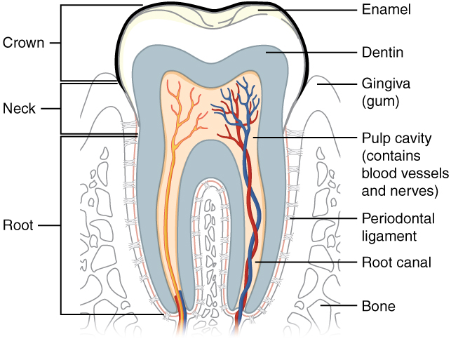
Trace the pulp and root canal to the apical region, then identify the periodontal ligament and supporting bone. Relate these structures to avulsion, apical pathology and extraction. The original uses the spelling “dentin”; cementum is not separately labelled.
Tooth & supporting tissues · OpenStax College · CC BY 3.0. Original image; displayed at a responsive size. Original publication.
Read the anatomy and predict resistance
Inspect the whole relevant image rather than only the crown. Assess root number, length, curvature, divergence, hypercementosis, previous root filling and surrounding radiolucency. Dense bone, ankylosis, a bulbous apex or divergent roots can make a small crown deceptively difficult. A two-dimensional overlap with the sinus or mandibular canal is not proof of direct contact.
Clinically assess access, mouth opening, crown strength, mobility, adjacent restorations and soft tissues. The maxillary tuberosity, mandibular canal, mental foramen and lingual tissues are potential injury sites. Imaging should be recent enough and sufficiently complete to answer the operative question; further exposure needs justification.
Adult mandible: labelled lateral anatomy
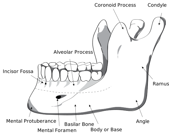
Locate the body, ramus, angle, condylar process, coronoid process and mental foramen. Use the labels to distinguish the two superior processes and the landmarks relevant to local anaesthesia and surgery.
HumanMandibleLeft.svg · Djexplo · CC0. None; reproduced without alteration.
Which nerve emerges through the mental foramen?
The mental nerve, a terminal branch of the inferior alveolar nerve, supplying the lower lip and chin.
Translate the medical history into a plan
Record medicines, allergies, previous bleeding and healing problems, antiresorptive exposure, radiotherapy and relevant systemic disease. Anticoagulation management depends on the medicine and procedure, with local haemostasis planned in advance; do not tell patients to stop treatment casually. Check the current SDCEP pathway rather than applying warfarin rules to a DOAC.
The setting must match anatomical difficulty, medical risk, anxiety, cooperation and available support. Local anaesthesia, sedation and general anaesthesia have different assessment and recovery requirements. A more complex anaesthetic does not compensate for an inadequate surgical plan or lack of competence.
Understand controlled luxation
Extraction separates periodontal ligament fibres and expands the socket sufficiently to deliver the root along a feasible path. Forceps need useful root purchase rather than a crushing grip on unsupported enamel. Movement should reflect root geometry: a conical root can behave differently from flattened or divergent roots.
Elevator force must be controlled and directed with the adjacent tooth and soft tissues protected. The neighbouring tooth is not a convenient fulcrum. Increasing resistance should prompt reassessment of purchase, access and anatomy. Planned sectioning or a surgical approach can reduce trauma when the original simple approach is no longer appropriate.
Respond safely to fracture or an unexpected finding
If the crown or root fractures, assess what remains under direct vision and with suitable localisation when needed. Consider size, mobility, infection, depth and proximity to a nerve or sinus before choosing retrieval. A deeply placed, small, non-infected fragment may sometimes be safer to retain than to pursue.
Intentional retention requires explanation, documentation and a review plan; it must not conceal a lost fragment. Stop blind exploration if a root is displaced or its location becomes uncertain. A suspected aspirated fragment changes the urgency and requires the appropriate emergency assessment.
Finish the procedure and plan recovery
Check the extracted tooth against the expected root anatomy and inspect the socket and neighbouring structures. Assess haemostasis, any sharp or diseased tissue and whether packing or sutures are needed. Avoid indiscriminate aggressive socket curettage. Record the tooth, technique, complications, fragments and measures used to control bleeding.
Give clear written and verbal advice on clot protection, hygiene, suitable analgesia, diet and smoking avoidance. Explain how to apply direct pressure for recurrent bleeding and where to seek help. Progressive swelling, difficulty swallowing or breathing, persistent bleeding and unexpected sensory change need a clear escalation route. Review depends on the procedure and the patient, rather than a universal appointment interval.
A distinction to remember
A tooth that is easy to remove can still be the wrong tooth to remove. Unexpected resistance should change the assessment before it changes the force.
A patient requests extraction of a painful lower molar, but the examination does not identify the source. What should happen first?
Reveal the reasoning
Establish a defensible diagnosis before irreversible treatment. Repeat the pain history, clinical tests and relevant imaging, including adjacent and referred causes. Explain the uncertainty and arrange appropriate further assessment; the patient’s request does not establish which tooth is responsible.
Test your recall
Which radiographic features can make an extraction difficult?
Show answer
Root curvature or divergence, bulbous apices, hypercementosis, ankylosis, dense surrounding bone and proximity to vital structures can increase difficulty. Interpret these alongside mouth opening, crown strength and access; a radiograph alone cannot establish the appropriate operator or setting.