
Oral biopsy
A biopsy is a diagnostic procedure with consequences for later treatment. Its value depends on selecting the right site, obtaining representative tissue, preserving it correctly and reconciling the report with the patient. A small incision is not automatically a low-risk or diagnostically simple operation.
Quick revision summary
Define the clinical question
Describe the lesion before choosing a technique: site, dimensions, colour, surface, ulceration, consistency, fixation and relation to adjacent anatomy. Record duration, progression, symptoms, medicines and systemic features. A clinical photograph or diagram helps the laboratory and future clinicians identify exactly which area was sampled.
Protect tissue and local structures
Plan anaesthesia, access, haemostasis and closure around ducts, nerves, vessels and underlying bone. Handle the sample gently; crush artefact and excessive heat can obscure epithelial architecture or margins. Electrosurgery or laser may be useful for selected tasks but can compromise a diagnostic edge if used without a suitable plan.
Close the diagnostic loop
Provide wound, bleeding and analgesic advice with an urgent contact route. Establish who will obtain, review and communicate the result and what happens if it is delayed. A missed result can be more harmful than an uncomplicated wound.
Common mistake
Formalin solves the routine histology question, but can destroy the value of a specimen intended for direct immunofluorescence. Plan the tests before taking tissue.
Oral biopsy: choosing, handling and interpreting a specimen
A biopsy is a diagnostic procedure with consequences for later treatment. Its value depends on selecting the right site, obtaining representative tissue, preserving it correctly and reconciling the report with the patient. A small incision is not automatically a low-risk or diagnostically simple operation.
Define the clinical question
Describe the lesion before choosing a technique: site, dimensions, colour, surface, ulceration, consistency, fixation and relation to adjacent anatomy. Record duration, progression, symptoms, medicines and systemic features. A clinical photograph or diagram helps the laboratory and future clinicians identify exactly which area was sampled.
Decide whether the question is inflammatory disease, dysplasia, malignancy, infection or another process. A suspected vascular lesion needs appropriate assessment before incision. Persistent or suspicious lesions should enter the relevant specialist or cancer pathway; arranging a routine local biopsy must not delay necessary referral.
Inflammation, granulation and maturation in tissue repair
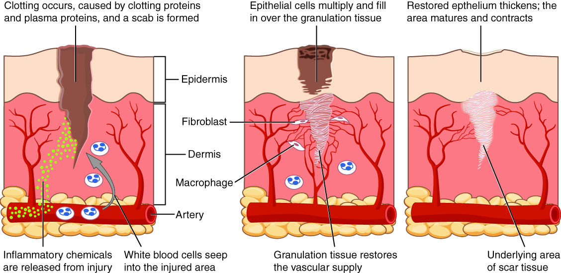
Relate inflammatory cells, fibroblasts, granulation tissue, epithelialisation and scar maturation to the overlapping stages of wound healing. This is a skin-wound example, not an extraction socket; oral mucosa differs and does not necessarily form a dry scab. Healing phases overlap.
Inflammation, granulation and maturation in tissue repair · OpenStax College (2013) · CC BY 3.0. Unchanged published image; displayed responsively. Original publication.
Choose incisional or excisional sampling
An incisional biopsy samples part of a lesion. It is often used for larger, heterogeneous or suspicious abnormalities where diagnosis guides definitive treatment. Include viable tissue and sufficient depth to demonstrate the relevant tissue architecture. Sampling only necrotic slough can miss the process responsible for the ulcer.
An excisional biopsy removes a selected lesion and may be appropriate for a small clinically benign abnormality when anatomy and closure are suitable. It is not permission to remove a suspected cancer casually with unplanned margins. Discuss the procedure’s aim, risks, limitations and the possibility of further treatment.
Select a representative area
The edge of an ulcer can capture abnormal epithelium and deeper tissue better than its necrotic centre, but the best location depends on the suspected disease. A large mixed red-white lesion may need more than one targeted sample, documented separately. An apparently bland area may not represent the highest-risk component.
For blistering disease, routine histology and direct immunofluorescence answer different questions. Specialist advice on an appropriate perilesional sample and transport medium should be obtained before the procedure. A fully eroded site can destroy the structures needed for interpretation; formalin is unsuitable for the routine DIF specimen.
Protect tissue and local structures
Plan anaesthesia, access, haemostasis and closure around ducts, nerves, vessels and underlying bone. Handle the sample gently; crush artefact and excessive heat can obscure epithelial architecture or margins. Electrosurgery or laser may be useful for selected tasks but can compromise a diagnostic edge if used without a suitable plan.
Use trained technique and equipment appropriate to the site. A scalpel, punch or other device is chosen for the question and anatomy, not convenience alone. Avoid tissue strangulation during closure. Where orientation matters, agree and record the marking system so that the pathologist can relate surfaces to the patient.
Match the specimen to the investigation
Routine histology commonly uses formalin according to the laboratory’s protocol. Microbiology, immunofluorescence and some other investigations need separate specimens and different handling. Contact the laboratory when uncertain before taking the tissue; an incorrectly fixed sample may not be recoverable for its intended test.
Label each container with patient identifiers and precise site, and complete the request with clinical history, differential, duration and relevant treatment. If there are multiple sites, make their identity unambiguous. Tissue for culture should not be treated as a superficial swab problem when a deeper sample is required.
Understand what the report can establish
Histology can define inflammatory patterns, dysplasia and invasion when the specimen captures them. A small incisional biopsy may not establish the worst grade across an entire heterogeneous lesion or the deepest extent. “No malignancy seen” describes the submitted tissue, not every unsampled area.
Read adequacy, diagnosis and limitations, and interpret margins according to whether the procedure was diagnostic or intended as a complete excision. An unexpected result or persistent clinical concern warrants discussion with the pathologist and specialist team. Clinical-pathological discordance is a reason to reassess, not an administrative inconvenience.
Close the diagnostic loop
Provide wound, bleeding and analgesic advice with an urgent contact route. Establish who will obtain, review and communicate the result and what happens if it is delayed. A missed result can be more harmful than an uncomplicated wound. Record the sample site and any intraoperative issues.
At review, assess healing and whether the result explains the lesion. Discuss next steps in understandable language, including further biopsy, treatment or surveillance. Ensure referral occurs through the appropriate urgency when malignancy or another significant diagnosis is found. A healed biopsy site does not itself demonstrate disease resolution.
A distinction to remember
Formalin solves the routine histology question, but can destroy the value of a specimen intended for direct immunofluorescence. Plan the tests before taking tissue.
A large red-white patch has one focal indurated area. Where should the biopsy plan focus?
Reveal the reasoning
Arrange appropriate specialist assessment and target representative abnormal tissue, including the clinically concerning component rather than sampling only an easy bland area. More than one site may be needed. Record each location clearly so pathology can be matched to the clinical findings.
Test your recall
How do incisional and excisional biopsies differ?
Show answer
An incisional biopsy samples part of a lesion for diagnosis; an excisional biopsy removes a selected lesion. Choice depends on the differential, size and anatomy. An incisional sample cannot ordinarily establish that the whole lesion has been removed or fully characterised.