
Pulp preservation
Pulp preservation retains living tissue where a suitable diagnosis, aseptic procedure and durable restoration make this realistic. Selective removal, pulp capping and pulpotomy are different treatment strategies.
Quick revision summary
Understand why preserving vitality matters
The pulp provides sensation, defence and dentine-forming capacity. In an immature permanent tooth, maintaining vitality can allow continued root development and strengthening of thin root walls. In mature teeth it also preserves biological functions and may avoid more extensive treatment.
Choose among exposed-pulp procedures
Direct pulp capping places a suitable material over an exposure without removing the coronal pulp. Partial pulpotomy removes a limited superficial portion, while full pulpotomy removes the coronal pulp to the canal orifices.
Plan surveillance and explain uncertainty
Review symptoms, function, restoration integrity, clinical tests and indicated radiographic findings. In immature teeth, assess continued development as well as absence of disease. After full pulpotomy, sensibility responses may be altered, so interpret them in relation to the tissue retained rather than using one absent cold response as proof of failure.
Common mistake
Pain labels and exposure size do not directly reveal the condition of the remaining pulp. Vital pulp treatment depends on the full case and an effective aseptic seal.
Pulp preservation
Pulp preservation retains living tissue where a suitable diagnosis, aseptic procedure and durable restoration make this realistic. Selective removal, pulp capping and pulpotomy are different treatment strategies.
Understand why preserving vitality matters
The pulp provides sensation, defence and dentine-forming capacity. In an immature permanent tooth, maintaining vitality can allow continued root development and strengthening of thin root walls. In mature teeth it also preserves biological functions and may avoid more extensive treatment. The aim is a comfortable, functional tooth with controlled disease, not simply avoiding root canal treatment at any cost.
Inflammation is not necessarily uniform throughout the pulp. Symptoms traditionally described as irreversible pulpitis do not precisely establish the histological condition of every part of the tissue. Contemporary guidance permits vital pulp treatment in selected symptomatic permanent teeth, but selection, asepsis and follow-up remain important. Do not convert this into a promise that any painful tooth can be capped.
Tooth & supporting tissues
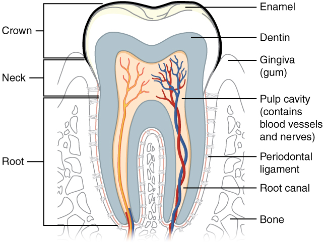
Trace the pulp and root canal to the apical region, then identify the periodontal ligament and supporting bone. Relate these structures to avulsion, apical pathology and extraction. The original uses the spelling “dentin”; cementum is not separately labelled.
Tooth & supporting tissues · OpenStax College · CC BY 3.0. Original image; displayed at a responsive size. Original publication.
Diagnose before deciding the intervention
Take a pain history including provocation, persistence, spontaneous episodes and night disturbance. Use sensibility tests with controls, percussion, palpation and indicated imaging. Assess cracks, restorability, caries depth, periodontal support and the ability to isolate and seal. A radiographic lesion reaching close to the pulp does not prove necrosis, and a symptom-free tooth is not automatically healthy.
Distinguish a vital pulp from a necrotic canal system and an apical condition. Trauma, immature roots, calcification and restorations can alter sensibility-test reliability. Record uncertainty when findings conflict and seek an appropriate assessment rather than committing to a procedure from one test. Consent should include realistic contingencies discovered during tissue removal.
The pulp–dentine interface
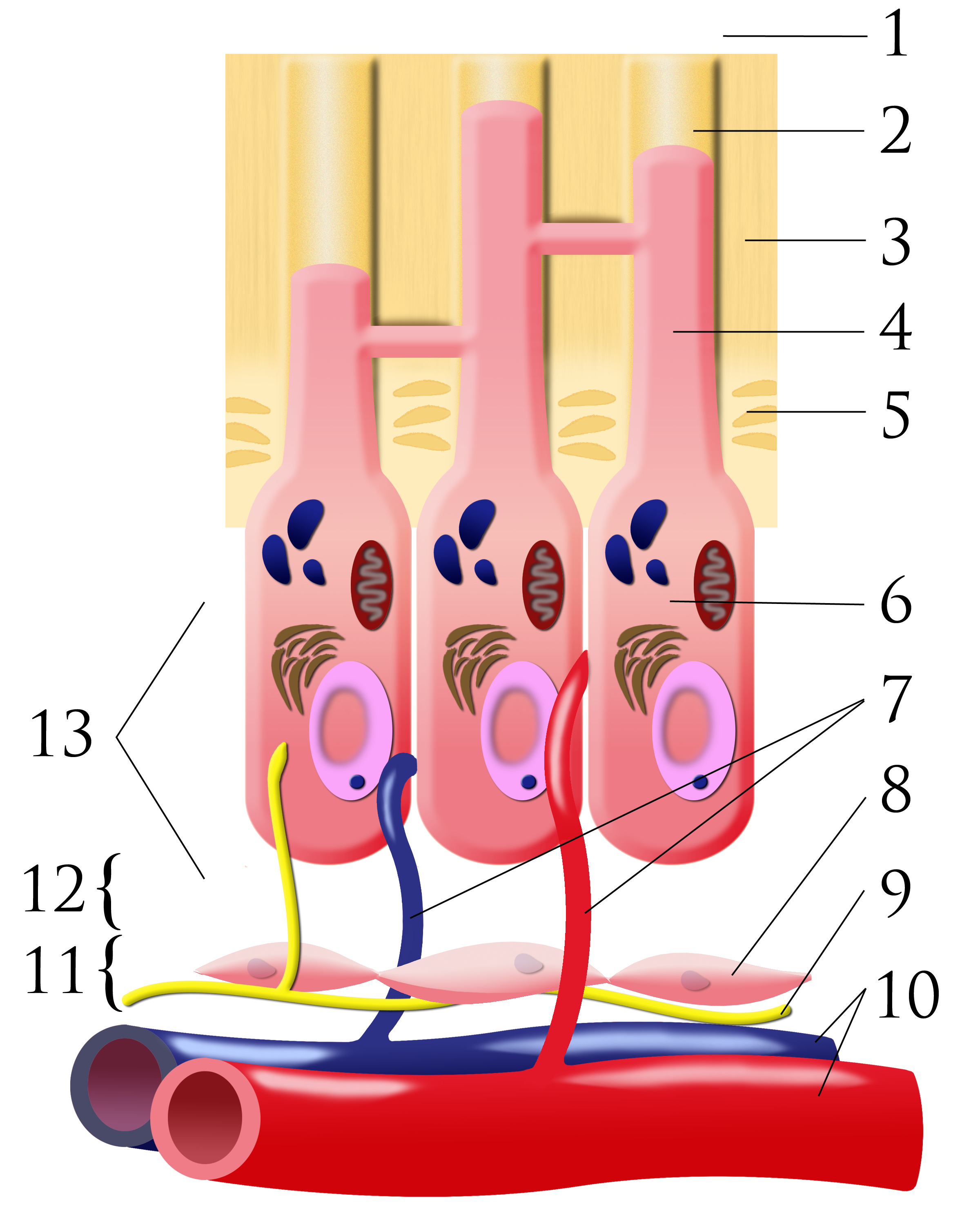
Number key: 1 outer-tooth direction; 2 dentinal tubule; 3 dentine; 4 odontoblastic process; 5 predentine; 6 odontoblast; 7 capillaries; 8 fibroblasts; 9 nerve; 10 artery and vein; 11 cell-rich zone; 12 cell-poor zone; 13 pulp chamber. This is a conceptual schematic, not a scale histological section.
Pulpal dentin junction.png · Ian Furst · CC BY-SA 3.0. None; original image reproduced without alteration.
Which cells form dentine, and where are their cell bodies?
Odontoblasts form dentine. Their cell bodies line the pulp–dentine border, with processes extending into dentinal tubules.
Preserve a dentine barrier when appropriate
In a deep carious lesion with a suitable pulpal diagnosis, selective removal may deliberately retain softer dentine near the pulp while establishing peripheral tissues suitable for a durable seal. The purpose is to reduce avoidable exposure and alter the lesion environment. It is a planned strategy, not an unsealed compromise or an instruction to leave all softened tissue everywhere.
A liner or protective material cannot compensate for a defective restoration. Choice depends on depth, substrate and the whole treatment plan. Explain whether the seal is intended to be definitive or whether re-entry is planned. An emergency temporary dressing and a formal stepwise strategy need different records and completion arrangements.
Choose among exposed-pulp procedures
Direct pulp capping places a suitable material over an exposure without removing the coronal pulp. Partial pulpotomy removes a limited superficial portion, while full pulpotomy removes the coronal pulp to the canal orifices. Pulpectomy removes pulp from the canal system and is part of root canal treatment. These names should describe what was done, not be used interchangeably.
Exposure context, contamination, tissue appearance, haemostasis, symptoms and restoration feasibility influence choice. A carious exposure is biologically different from a fresh traumatic or mechanical exposure. Size alone is not a reliable treatment rule. If findings suggest that the intended remaining tissue is unsuitable, reassess rather than placing a material merely to finish the planned procedure.
| Procedure | Tissue approach | Key distinction |
|---|---|---|
| Selective removal | Dentine barrier intentionally retained near pulp | No intended pulp exposure |
| Direct capping | Material over exposed pulp | No coronal pulp removal |
| Partial pulpotomy | Superficial pulp removed | Retains more coronal pulp |
| Full pulpotomy | Coronal pulp removed | Radicular tissue retained |
| Pulpectomy | Canal pulp removed | Root canal treatment pathway |
Asepsis, material and coronal seal work together
Use dental dam and a controlled field, good illumination and appropriate magnification. Contemporary vital pulp procedures commonly use hydraulic calcium-silicate materials with product-specific indications and handling. Select the material and restoration as a compatible plan, including the risk of discolouration where relevant. A material marketed as bioactive does not establish that every pulp or exposure is suitable.
Haemostasis and tissue assessment inform the procedure, but avoid applying an arbitrary timer as the sole diagnosis. Use current guidance and the operator’s training for the technique. Achieve a prompt durable coronal seal and adequate structural protection. Persistent contamination or inability to restore the tooth changes the likelihood that tissue preservation can succeed.
Plan surveillance and explain uncertainty
Review symptoms, function, restoration integrity, clinical tests and indicated radiographic findings. In immature teeth, assess continued development as well as absence of disease. After full pulpotomy, sensibility responses may be altered, so interpret them in relation to the tissue retained rather than using one absent cold response as proof of failure.
Warn about possible persistent pain, necrosis, apical disease, discolouration and later need for root canal treatment or extraction. A successful immediate seal does not prove long-term pulp health. Define who will review, how deterioration should be reported and what outcome is expected. Refer when case complexity or the necessary technique exceeds available competence or equipment.
A distinction to remember
Pain labels and exposure size do not directly reveal the condition of the remaining pulp. Vital pulp treatment depends on the full case and an effective aseptic seal.
A mature molar has spontaneous pain but remains vital. Is root canal treatment the only possible tooth-retaining option?
Reveal the reasoning
Not necessarily. ESE guidance allows full pulpotomy as an option in selected permanent teeth as well as root canal treatment. Diagnosis, operative tissue findings, asepsis, restorative prognosis and operator competence determine suitability. Discuss uncertainties and the possibility of later treatment rather than promising equivalence in every case.
Test your recall
How does full pulpotomy differ from pulpectomy?
Show answer
Full pulpotomy removes the coronal pulp while retaining radicular tissue; pulpectomy removes pulp from the root canal system as part of root canal treatment. The retained tissue, biological aim and follow-up differ. Record the actual procedure rather than using the names interchangeably.