
CBCT
Three-dimensional information brings additional responsibilities as well as potential benefit.
Quick revision summary
Justify the three-dimensional question
Identify the question that two-dimensional imaging has not resolved and how its answer would change management. Examples include selected anatomical relationships or complex diagnostic situations; the indication must be individual.
Balance useful detail against exposure and artefact
Field of view determines how much anatomy is acquired; voxel size contributes to spatial resolution but does not alone determine diagnostic quality. Movement, noise and metal artefact can obscure the target despite a small voxel.
Interpret the complete acquired volume
Review multiplanar information with the necessary training rather than relying on a single attractive reconstruction. Artefacts around metal can mimic or hide defects, and limited soft-tissue contrast makes CBCT unsuitable for some questions.
Common mistake
A larger or more detailed scan is not automatically a better investigation.
CBCT selection and full-volume interpretation
Three-dimensional information brings additional responsibilities as well as potential benefit.
Justify the three-dimensional question
Identify the question that two-dimensional imaging has not resolved and how its answer would change management. Examples include selected anatomical relationships or complex diagnostic situations; the indication must be individual. Choose an appropriate field of view, resolution and exposure protocol. Greater resolution or a larger volume is not automatically better if it adds exposure or incidental findings without benefit.
CBCT has artefacts, including those associated with metal and movement, and limited soft-tissue characterisation. The complete acquired volume must be interpreted by someone with appropriate competence, with specialist reporting where necessary. Do not assess only a few familiar slices around the target tooth. Record the report and integrate its findings into consent, treatment planning and any incidental-finding pathway.
Stafne bone cavity: the lingual depression on CT
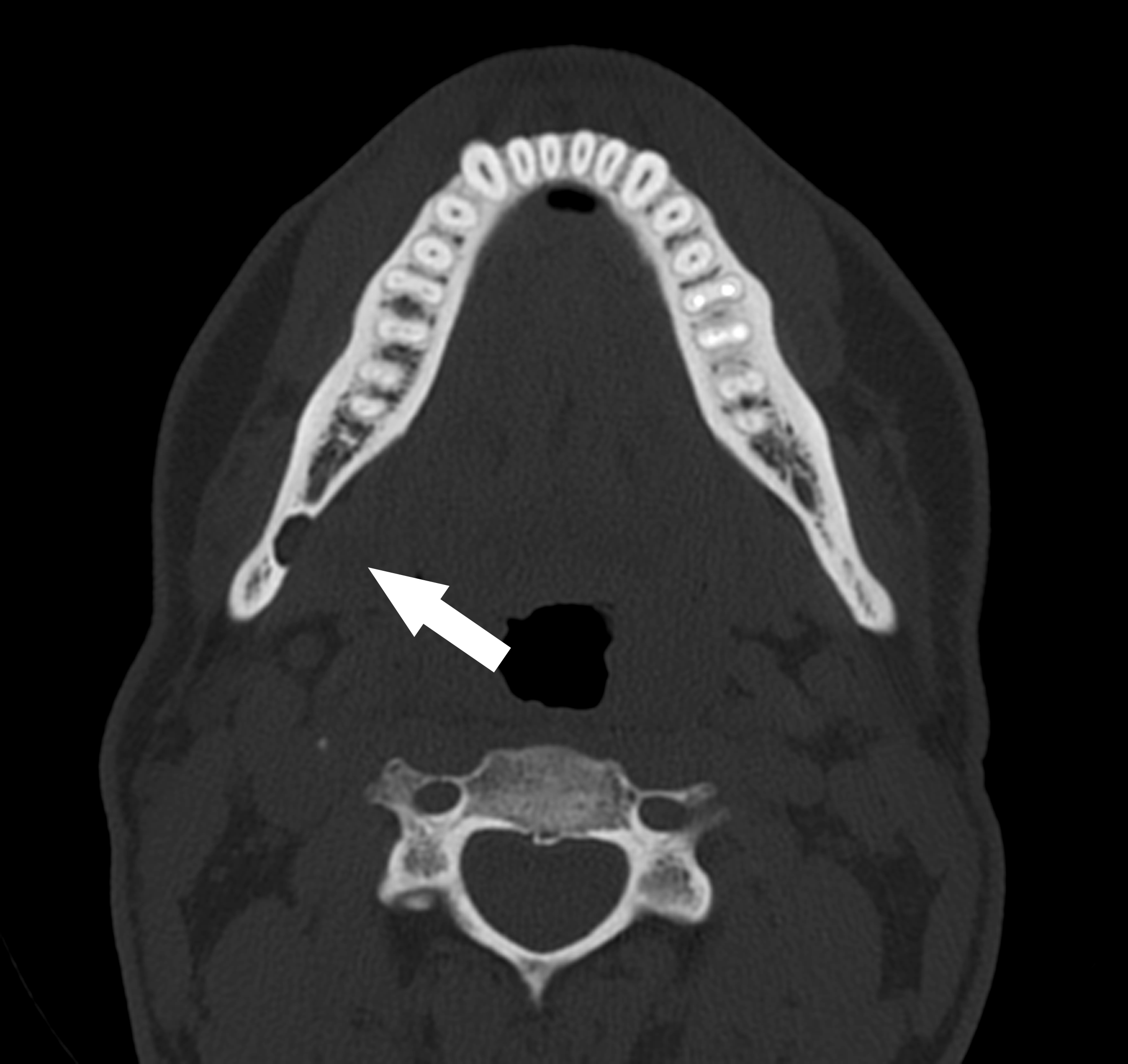
The original arrow marks a concavity in the lingual mandibular cortex. Compare it with the panoramic example of the same source case. This helps explain why a two-dimensional radiolucency can represent a bony depression rather than an epithelial-lined odontogenic cyst. Further imaging in a real case needs an individual clinical indication.
Stafne defect CT axial.jpg · Coronation Dental Specialty Group · CC BY 3.0. Reproduced without alteration. Labels, arrows and circles, where present, belong to the source image.
Why should a Stafne bone cavity not automatically be called a true odontogenic cyst?
It represents a mandibular bony depression rather than an epithelial-lined tooth-derived cyst. Classification depends on the complete assessment.
Key distinctions in practice
| Finding or option | What it means | Clinical consequence |
|---|---|---|
| Clinical question | Information likely to change a decision | Justify against existing and lower-exposure alternatives. |
| Field of view | Extent of the acquired volume | Use appropriate coverage rather than routine maximum size. |
| Artefact | A source of unreliable appearance | Recognise limits around metal or movement. |
| Full-volume report | Responsibility for all acquired information | Arrange competent interpretation and action. |
Balance useful detail against exposure and artefact
Field of view determines how much anatomy is acquired; voxel size contributes to spatial resolution but does not alone determine diagnostic quality. Movement, noise and metal artefact can obscure the target despite a small voxel. Increasing detail or field size without a clinical benefit adds burden rather than certainty.
Choose the smallest appropriate field and a protocol adequate for the question through the trained imaging team. Consider whether an existing image or a suitable lower-exposure examination already answers it. A CBCT should not be a routine screening exercise or an automatic part of every endodontic, surgical or implant assessment. The expected effect on the decision is central to justification.
Interpret the complete acquired volume
Review multiplanar information with the necessary training rather than relying on a single attractive reconstruction. Artefacts around metal can mimic or hide defects, and limited soft-tissue contrast makes CBCT unsuitable for some questions. A negative scan therefore needs interpretation in relation to the suspected condition and the scan’s limitations.
Arrange reporting by someone competent for the entire volume, with radiology support for anatomy or abnormalities outside the operator’s expertise. Record significant incidental findings and ensure a follow-up pathway. The scan can clarify an anatomical relationship, but it cannot select the treatment on its own; combine its findings with indication, alternatives, patient priorities and procedural risk.
A distinction to remember
A larger or more detailed scan is not automatically a better investigation.
If CBCT is taken for one implant site, can the remaining volume be ignored?
Reveal the reasoning
No. The complete acquired volume requires appropriate interpretation and an action pathway for relevant incidental findings.
Guidance to read alongside this chapter
Test your recall
Does a CBCT automatically replace conventional images?
Show answer
No; the added information must justify the higher exposure.