
Periodontal prevention
Periodontal prevention combines daily biofilm control, modification of susceptibility and surveillance. A treated patient remains someone with a history of periodontitis, even when inflammation is controlled.
Quick revision summary
Gingivitis and periodontitis are different endpoints
Plaque-associated gingivitis is inflammation without attachment loss attributable to periodontitis. It can resolve with effective plaque control. Periodontitis involves loss of the supporting attachment apparatus and bone in a susceptible host.
Teach a precise daily routine
Show the patient where plaque persists and demonstrate a brushing method that reaches the gingival margins and the final teeth. Select interdental aids by space, anatomy and dexterity, using return demonstration.
Explain prognosis without blame
Describe which teeth or sites are most vulnerable and why, separating current inflammation from historical loss. Give the patient a practical role in the plan and acknowledge barriers such as dexterity, cost, competing illness or care dependence.
Common mistake
BPE cannot monitor a known periodontitis patient in place of appropriate detailed charting, and a scale alone is not a preventive plan.
Periodontal prevention
Periodontal prevention combines daily biofilm control, modification of susceptibility and surveillance. A treated patient remains someone with a history of periodontitis, even when inflammation is controlled.
Gingivitis and periodontitis are different endpoints
Plaque-associated gingivitis is inflammation without attachment loss attributable to periodontitis. It can resolve with effective plaque control. Periodontitis involves loss of the supporting attachment apparatus and bone in a susceptible host. Treatment can stabilise the disease, but it does not return the tooth to an untouched periodontium. This distinction explains why a previously treated patient needs continuing supportive care.
Visible inflammation alone does not describe past destruction or future risk. Recession, attachment level, pocketing and bone support need interpretation together. A reduced periodontium may be stable, while a patient with little visible plaque can still have an important neglected site. Smoking and other factors can alter the clinical appearance, so examine rather than infer health from a single sign.
Gingivitis: inflamed gingiva without attachment loss
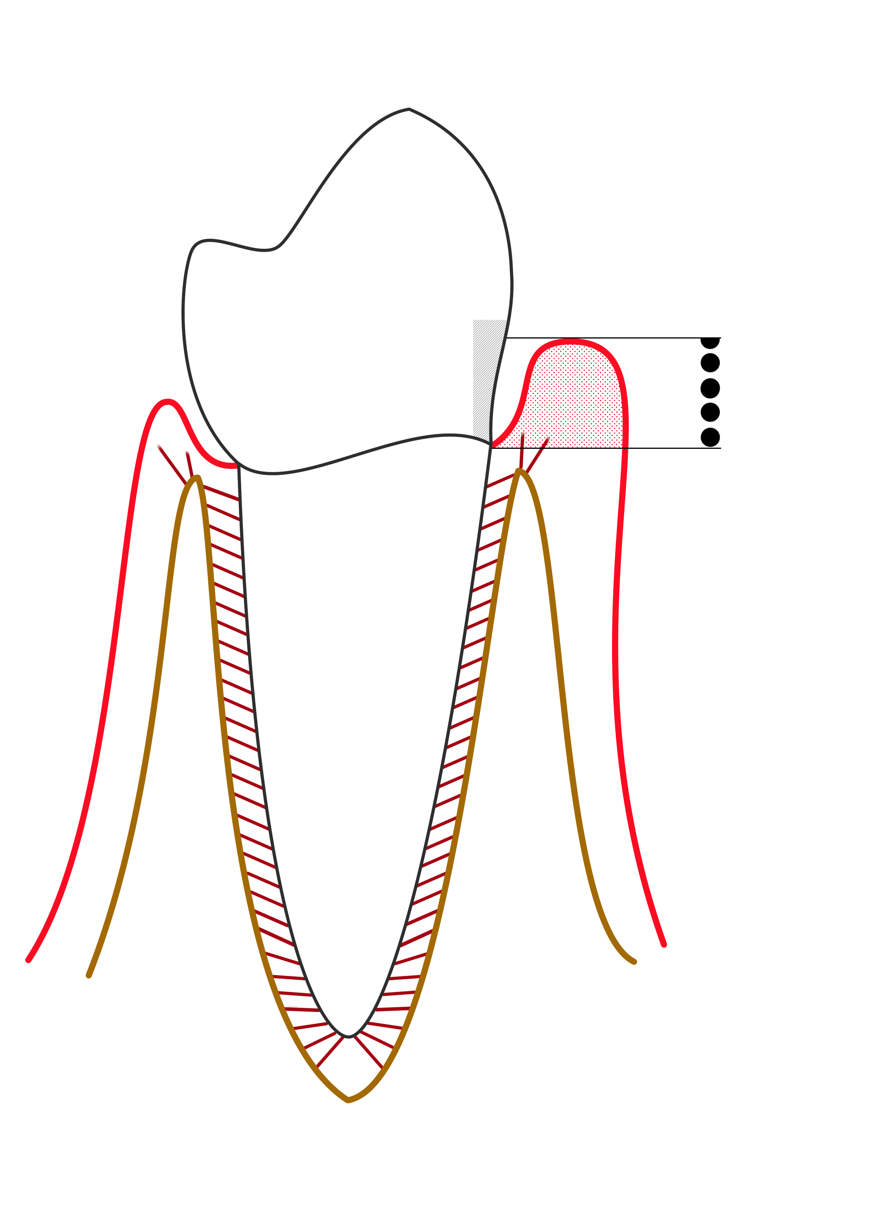
The red marginal outline represents gingival inflammation. Compare the gingival margin, sulcus and supporting bone with the periodontitis diagram. The probe-scale dots illustrate depth, not a diagnosis by themselves.
Line diagram of Gingivitis.png · Ian Furst · CC BY-SA 4.0. None; reproduced without alteration.
Does gingival bleeding alone establish periodontitis?
No. Periodontitis requires evidence of supporting attachment loss, interpreted with clinical and radiographic findings.
Screen appropriately, then investigate
BPE is a screening tool, not a diagnosis or a detailed follow-up chart. SDCEP recommends it for new adults and adults without a periodontitis diagnosis at recall. A history of periodontitis calls for appropriate full periodontal examination rather than replacing this with another BPE. Record the highest sextant score and furcation involvement while examining all relevant sites.
Current SDCEP advice requires further assessment for BPE 3, 4 or a furcation marker. For code 3, chart the affected sextant after initial treatment; code 4 requires whole-mouth charting before treatment. Interpret findings with attachment loss, indicated radiographs, risk and causes of non-periodontal recession. A pocket measurement alone does not stage or grade the patient.
Periodontitis: pocket formation and loss of support
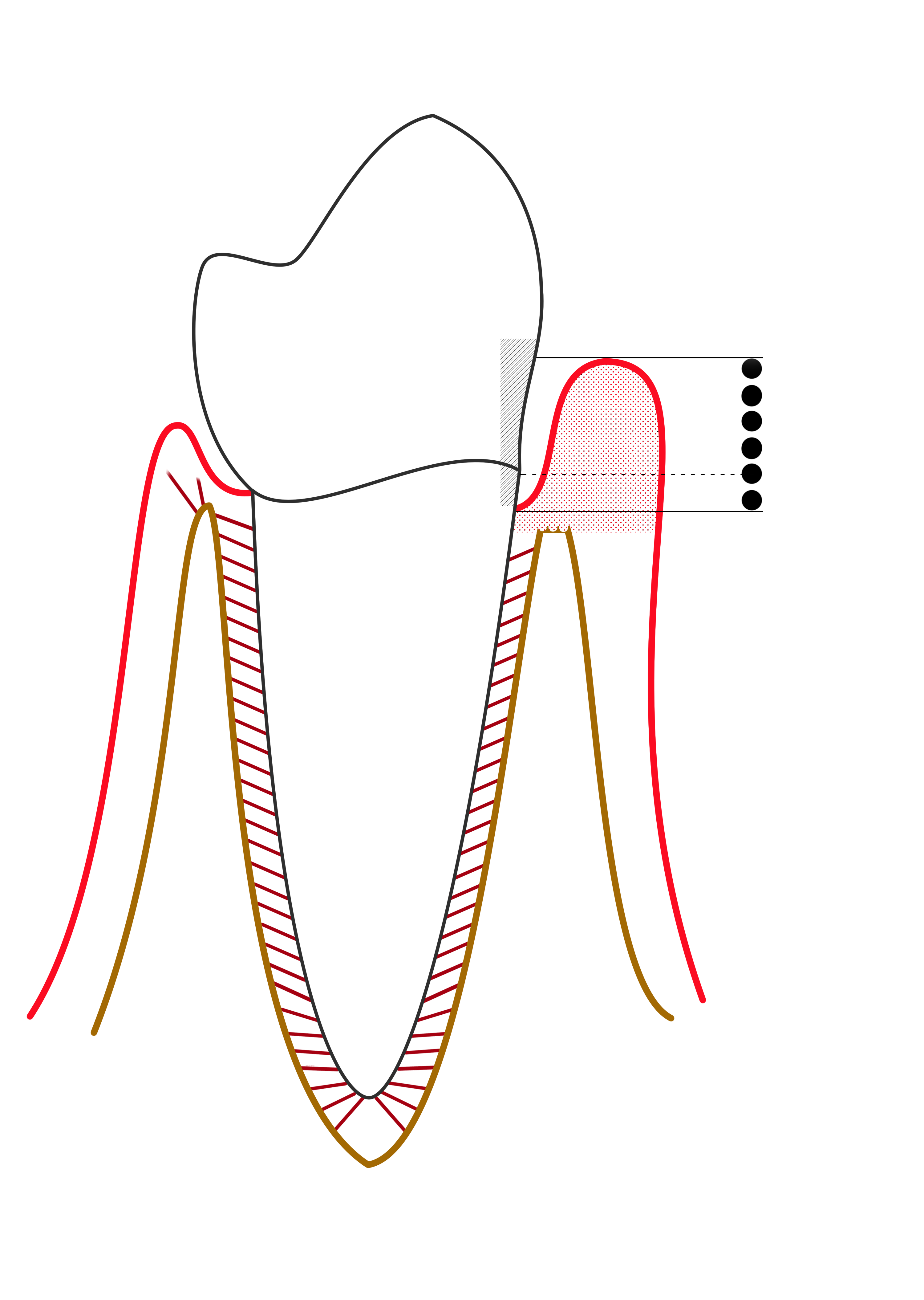
Compare the apical extent of the pocket and the reduced supporting bone on the affected side. This simplified diagram is labelled “mild periodontitis” by its author; it is not a modern stage/grade classification chart.
Mild periodontitis.png · Ian Furst · CC BY-SA 4.0. None; reproduced without alteration.
Why is probing depth different from clinical attachment level?
Probing depth runs from the gingival margin to the pocket base. Clinical attachment level uses the cemento-enamel junction as its reference; recession or enlargement changes their relationship.
Identify the risks that can be changed
Daily plaque control is essential but does not fully explain variation in disease susceptibility. Ask about smoking, diabetes and its management, relevant medicines, previous progression and attendance barriers. Discuss glycaemic control sensitively and coordinate with medical care where needed; do not offer dental treatment as a replacement for diabetes management or promise a particular blood-test improvement.
Local risks include calculus, restoration overhangs, open margins, inaccessible contacts and plaque-retentive prostheses. Correct these where feasible. A complex crown or bridge should be designed for maintenance in the patient’s actual mouth. If cleaning is already difficult, adding a bulky contour can create a preventable recurrence site despite otherwise successful treatment.
Teach a precise daily routine
Show the patient where plaque persists and demonstrate a brushing method that reaches the gingival margins and the final teeth. Select interdental aids by space, anatomy and dexterity, using return demonstration. Bleeding can cause avoidance, so explain the purpose of gentle cleaning and distinguish inflammation from traumatic technique. A small, achievable change is a useful beginning when inflammation is extensive.
Support smoking cessation and agree goals linked to the patient’s priorities. Professional supra- and subgingival instrumentation is selected according to diagnosis and need; it complements home care. A routine polish or mouthrinse is not an equivalent treatment for untreated periodontitis. Reassess the tissue response before deciding that a patient has reached a maintenance phase.
Understand supportive periodontal care
After active therapy, review symptoms, plaque, bleeding, pocketing, attachment findings and relevant risk changes. Compare with a meaningful baseline and use site-specific charting as appropriate. Stability requires more than the absence of pain, and a periodontitis patient is not reclassified as never having had the disease simply because they now clean well.
Tailor supportive-care frequency to disease severity, response, recurrence risk and ability to maintain the result. This interval differs from a routine dental examination interval. Re-instrumentation, reinforcement of self-care and correction of local factors may be needed at selected sites. Deep residual or recurrent pockets, persistent bleeding and further attachment loss should trigger reassessment and consideration of referral.
| Situation | What remains important | Review approach |
|---|---|---|
| Gingivitis | Biofilm and reversible inflammation | Check cleaning and resolution |
| Untreated periodontitis | Attachment loss and active disease | Diagnosis-based active treatment |
| Treated stable periodontitis | Reduced support and continuing susceptibility | Individual supportive care |
| Recurrent local inflammation | Site-specific cause or progression | Investigate access and chart the site |
Explain prognosis without blame
Describe which teeth or sites are most vulnerable and why, separating current inflammation from historical loss. Give the patient a practical role in the plan and acknowledge barriers such as dexterity, cost, competing illness or care dependence. Continued disease is a reason to investigate and adapt the plan, not merely record that the patient is non-compliant.
At review, ask what was achieved and what remained difficult. A favourable average plaque score can hide a failing molar furcation or an inaccessible distal site. Document the management of that site and the next review target. Prevention is successful when the supporting tissues remain stable and the routine can be sustained, not when a predetermined cleaning appointment has been completed.
A distinction to remember
BPE cannot monitor a known periodontitis patient in place of appropriate detailed charting, and a scale alone is not a preventive plan.
A patient previously treated for periodontitis has little bleeding and no pain. Why should maintenance continue?
Reveal the reasoning
The reduced support and history of susceptibility remain relevant despite a good response. Assess site-specific probing and attachment findings, plaque access and risks, then tailor supportive care. Absence of pain or a low bleeding observation alone cannot establish long-term stability.
Guidance to read alongside this chapter
Test your recall
What separates plaque-associated gingivitis from periodontitis?
Show answer
Gingivitis is inflammation without attachment loss attributable to periodontitis and can resolve with effective plaque control. Periodontitis involves destruction of the supporting apparatus. A successfully treated patient may be stable but retains the diagnosis and maintenance needs associated with that history.