
Physiology and healing
Physiology explains how normal oral functions are maintained and how tissue responds to injury. Saliva, sensation, perfusion, haemostasis and mechanical stability all influence what a patient experiences during recovery.
Quick revision summary
Homeostasis, perfusion and oxygen delivery
Homeostasis maintains internal conditions through feedback, including regulation of temperature, glucose and fluid balance. Tissue oxygen delivery depends on blood flow, haemoglobin concentration and oxygen saturation, not saturation alone. Local perfusion also determines delivery of immune cells, nutrients and many medicines.
Haemostasis creates the initial repair scaffold
Vessel injury triggers vasoconstriction, platelet adhesion, activation and aggregation. Coagulation generates thrombin and a fibrin network that stabilises the initial platelet plug. Fibrinolysis subsequently contributes to clot removal as repair proceeds.
Connect impaired healing to a mechanism
Smoking, poor glycaemic control, malnutrition, ischaemia, infection and some treatments impair different parts of repair. Tension can reduce edge perfusion, dead space can collect fluid, and thermal or crush injury can leave devitalised tissue.
Common mistake
A closed mucosal surface does not establish mature collagen strength, complete socket mineralisation or restored ridge volume. Granulation tissue and granulomatous inflammation are different processes.
Physiology and healing
Physiology explains how normal oral functions are maintained and how tissue responds to injury. Saliva, sensation, perfusion, haemostasis and mechanical stability all influence what a patient experiences during recovery.
Homeostasis, perfusion and oxygen delivery
Homeostasis maintains internal conditions through feedback, including regulation of temperature, glucose and fluid balance. Tissue oxygen delivery depends on blood flow, haemoglobin concentration and oxygen saturation, not saturation alone. Local perfusion also determines delivery of immune cells, nutrients and many medicines. A normal-looking wound surface does not measure these processes directly.
Inflammation increases local blood flow and vascular permeability, producing warmth, redness and oedema. Some inflammation is part of repair, but impaired perfusion, ongoing infection or excessive tissue pressure can compromise recovery. A poorly perfused flap may fail despite technically neat sutures because closing the edges does not replace its vascular supply.
Stages of secondary bone healing
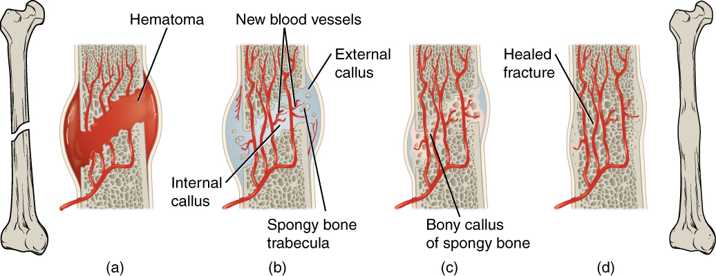
Follow haematoma, callus formation and remodelling. This general secondary-fracture-healing model uses a long bone; it is not an extraction-socket diagram or a depiction of primary healing under rigid fixation.
Stages of secondary bone healing · OpenStax College · CC BY 3.0. Original image; displayed at a responsive size. Original publication.
Saliva and swallowing are active physiological systems
Acinar cells produce salivary fluid and proteins; ducts modify the secretion. Both autonomic divisions influence secretion, with parasympathetic activity strongly promoting a plentiful watery flow. Saliva lubricates, supports taste and swallowing, buffers acids, supplies calcium and phosphate and contributes antimicrobial defence. Its importance is chemical as well as mechanical.
Xerostomia is the subjective sensation of dryness, whereas hyposalivation is a measured reduction in secretion; the two overlap without being identical. Medicines, gland disease, dehydration and head-and-neck radiotherapy may contribute. Reduced lubrication and buffering can produce discomfort, dysphagia, candidosis and rapidly progressing caries. Swallowing requires coordinated tongue propulsion, pharyngeal movement and airway protection rather than a single muscle action.
Inflammation, granulation and maturation in tissue repair
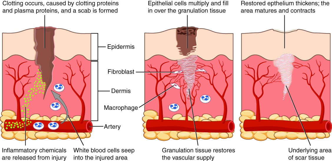
Relate inflammatory cells, fibroblasts, granulation tissue, epithelialisation and scar maturation to the overlapping stages of wound healing. This is a skin-wound example, not an extraction socket; oral mucosa differs and does not necessarily form a dry scab. Healing phases overlap.
Inflammation, granulation and maturation in tissue repair · OpenStax College (2013) · CC BY 3.0. Unchanged published image; displayed responsively. Original publication.
Pain, nociception and altered sensation
Nociceptors detect potentially damaging stimuli and transmit signals through peripheral and central pathways. Tissue mediators can lower their activation threshold, producing tenderness around injury. Pain also depends on central processing, previous experience and context; severity is not a direct measure of tissue destruction.
Dentine sensitivity can involve hydrodynamic movement in tubules. Neuropathic pain instead results from a lesion or disease of the somatosensory system and may include burning, electric shocks, allodynia or altered sensation. Allodynia means pain from a normally non-painful stimulus; hyperalgesia is an exaggerated response to a painful stimulus. These descriptions help assess persistent pain without automatically assigning a dental source.
Haemostasis creates the initial repair scaffold
Vessel injury triggers vasoconstriction, platelet adhesion, activation and aggregation. Coagulation generates thrombin and a fibrin network that stabilises the initial platelet plug. Fibrinolysis subsequently contributes to clot removal as repair proceeds. Platelet function and coagulation are related but distinct, which helps explain why different medicines or disorders produce different bleeding patterns.
A socket clot is a temporary biological scaffold. Gentle tissue handling and effective local haemostasis protect it; excessive disruption, a persistent source of bleeding or infection can interfere with healing. Local pressure and appropriate haemostatic measures work at the wound, while decisions about antithrombotic medicines require the relevant procedure-specific guidance.
The overlapping phases of soft-tissue repair
Neutrophils dominate early clearance after injury, while macrophages remove debris and coordinate later repair. During proliferation, capillaries grow, fibroblasts deposit matrix and epithelial cells migrate over the surface. Myofibroblasts contribute contraction. Granulation tissue is this vascular reparative tissue; it is different from a granuloma, which is a macrophage-based inflammatory pattern.
Remodelling reorganises collagen, changes its composition and improves mechanical strength over time. Epithelial closure occurs before full connective-tissue maturation, and scarred tissue does not necessarily regain the original architecture. The phases overlap rather than switching abruptly on a fixed postoperative day.
| Healing pattern | What happens | Practical consequence |
|---|---|---|
| Primary intention | Approximated viable edges heal across a small gap | Requires suitable tissue, perfusion and manageable tension |
| Secondary intention | A defect fills with granulation tissue and contracts | More tissue replacement and surface migration are needed |
| Delayed primary closure | Closure follows an initial period of wound management | Used when immediate closure is unsuitable |
| Remodelling | Matrix is reorganised after initial closure | Appearance and mature mechanical strength have different timelines |
Socket healing and fracture repair
An extraction socket progresses from clot to granulation tissue and then increasing bone formation and remodelling. Surface mucosa can close while the socket remains incompletely mineralised. Ridge width and height change after extraction because healing includes resorption and remodelling; a closed socket therefore does not imply preservation of the original alveolar dimensions.
Secondary fracture healing passes through haematoma, soft callus, hard callus and remodelling when relative stability and perfusion permit. Direct bone healing can occur with very high stability and close apposition with little external callus. Mechanical instability, a gap, poor blood supply or infection can impair union. A long-bone callus illustration explains this mechanism but is not a literal picture of socket healing.
Connect impaired healing to a mechanism
Smoking, poor glycaemic control, malnutrition, ischaemia, infection and some treatments impair different parts of repair. Tension can reduce edge perfusion, dead space can collect fluid, and thermal or crush injury can leave devitalised tissue. Simply tying a suture tighter may worsen these problems.
Review recovery against the procedure and expected course. Escalating pain, discharge, wound separation, exposed abnormal bone or persistent neurological change need assessment of their cause. Do not attribute every delay to a background risk factor while overlooking a local problem. Good follow-up identifies both the biological issue and an achievable response.
A distinction to remember
A closed mucosal surface does not establish mature collagen strength, complete socket mineralisation or restored ridge volume. Granulation tissue and granulomatous inflammation are different processes.
A flap is tightly sutured but its edge becomes pale and later separates. What mechanism should be considered?
Reveal the reasoning
Excessive closure tension may have reduced perfusion, with ischaemia and breakdown despite close initial approximation. Review flap design, vascular supply, tissue trauma and infection. A tighter replacement suture cannot compensate for a poorly perfused or inadequately mobilised flap.
Guidance to read alongside this chapter
Test your recall
What determines oxygen delivery to a healing tissue?
Show answer
Blood flow, haemoglobin concentration and haemoglobin oxygen saturation all contribute. Saturation alone cannot establish adequate delivery. This is why impaired perfusion or anaemia can matter to healing even when a pulse oximeter gives a satisfactory reading.