
General pathology
Pathology describes how cells and tissues adapt, become injured and produce disease. Distinguish a mechanism, a tissue pattern and a final diagnosis: they are different levels of explanation.
Quick revision summary
Cell adaptation and reversible injury
Hypertrophy increases cell size, while hyperplasia increases cell number. Atrophy reduces tissue size through smaller or fewer cells. Metaplasia replaces one mature cell type with another in response to an altered environment.
Circulation, fluid imbalance and tissue damage
Oedema is excess interstitial fluid. It can result from increased hydrostatic pressure, reduced plasma oncotic pressure, increased vascular permeability or impaired lymphatic drainage. Local inflammatory oedema and dependent oedema from systemic disease therefore have different mechanisms.
Specimens, clinical correlation and diagnostic uncertainty
An incisional biopsy samples part of a lesion; an excisional biopsy attempts to remove it. Site selection, depth, handling and the information supplied affect interpretation. For suspected immune blistering disease, a specimen for immunofluorescence may require different site and transport from routine histology.
Common mistake
Dysplasia, invasion, differentiation and stage answer different questions. A reassuring sample cannot be interpreted independently of whether it represents the clinically important part of the lesion.
General pathology
Pathology describes how cells and tissues adapt, become injured and produce disease. Distinguish a mechanism, a tissue pattern and a final diagnosis: they are different levels of explanation.
Cell adaptation and reversible injury
Hypertrophy increases cell size, while hyperplasia increases cell number. Atrophy reduces tissue size through smaller or fewer cells. Metaplasia replaces one mature cell type with another in response to an altered environment. These adaptations may be physiological or pathological and are not automatically malignant.
Hypoxia, toxins, physical injury, infection and immune injury can disrupt energy production, membranes and protein handling. Early injury may be reversible if the cause is removed. The threshold for irreversible damage depends on the cell type, duration and severity of the insult. The same stimulus can therefore produce adaptation in one setting and cell death in another.
Oral mucosa: epithelium, basement membrane and lamina propria
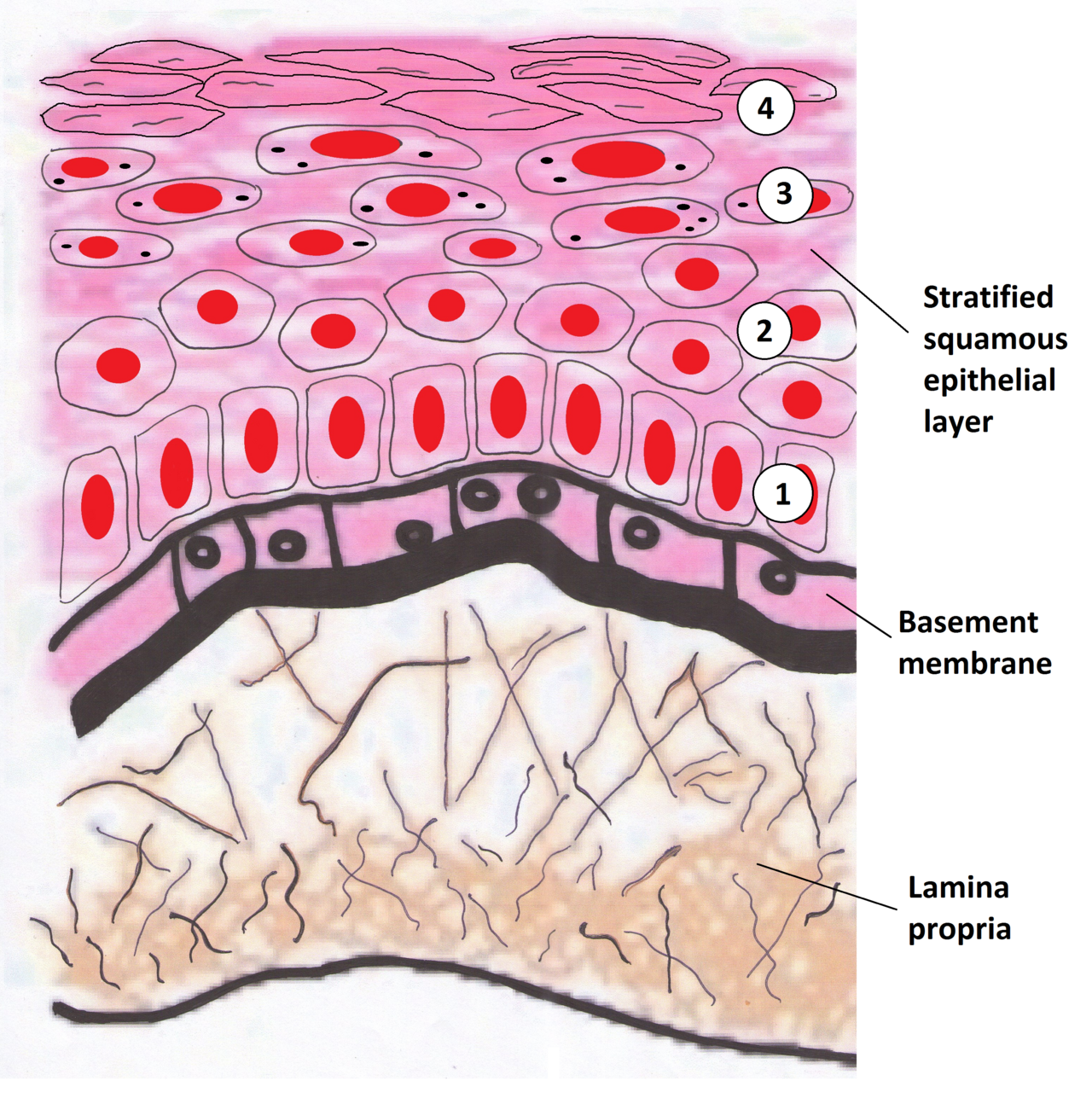
Identify the epithelium, basement membrane and lamina propria. Relate the epithelial–stromal interface to in-situ disease and invasion. This drawing represents a keratinising surface; oral sites have different keratinisation patterns. Use the printed anatomical names for revision.
Oral mucosa: epithelium, basement membrane and lamina propria · Wiki-minor (own work, 2011) · CC BY-SA 3.0. Unchanged published raster preview; displayed responsively. Original available from the source.
Necrosis, apoptosis and the surrounding reaction
Necrosis involves loss of membrane integrity and release of cellular contents, generally provoking inflammation. Its tissue pattern depends on the cause and site. Apoptosis is regulated cellular dismantling with removal of fragments, often producing less surrounding inflammation. It is important in normal development and tissue turnover as well as disease.
An ulcer describes loss of surface epithelium exposing underlying tissue; erosion is a more superficial epithelial defect. Neither term establishes the cause. A clinical ulcer can arise from trauma, infection, immune disease or malignancy, so it must be described by site, size, morphology, duration and associated findings before it is assigned an aetiology.
Acute inflammation, chronic inflammation and repair
Acute inflammation involves vascular change, fluid exudation and cellular recruitment, often with neutrophils prominent. Chronic inflammation commonly contains macrophages and lymphocytes together with tissue destruction and attempted repair. These are patterns rather than strict calendar definitions; the underlying stimulus determines whether inflammation resolves, persists or progresses.
An abscess is a localised pus collection. Granulation tissue is vascular tissue involved in repair, whereas granulomatous inflammation consists of organised activated macrophages, sometimes with giant cells. A granuloma can reflect infection, foreign material or inflammatory disease. Calling a specimen “granulomatous” does not identify one exclusive cause.
Circulation, fluid imbalance and tissue damage
Oedema is excess interstitial fluid. It can result from increased hydrostatic pressure, reduced plasma oncotic pressure, increased vascular permeability or impaired lymphatic drainage. Local inflammatory oedema and dependent oedema from systemic disease therefore have different mechanisms. A swelling may also be a solid mass or a fluid-filled lesion rather than oedema.
A thrombus forms within a vessel or the heart during life; an embolus travels and lodges elsewhere. Ischaemia is inadequate blood supply, whereas infarction is tissue necrosis caused by impaired perfusion. Congestion reflects impaired venous outflow. These distinctions explain why both arterial compromise and venous obstruction can threaten tissue viability, although their appearances and management differ.
Neoplasia, dysplasia and invasion
A neoplasm is an abnormal clonal growth with biological behaviour that requires classification. Benign lesions do not metastasise but can still cause serious effects through size, site, secretion or local destruction. Malignancy involves invasive and potentially metastatic behaviour; speed of growth, pain or surface colour alone cannot reliably classify every lesion.
Epithelial dysplasia includes disordered architecture and cytological atypia. Invasive epithelial cancer has crossed the basement-membrane boundary into underlying tissue. Differentiation describes resemblance to the tissue of origin. A clinical white patch is not itself a dysplasia grade, and an apparently small lesion can contain important microscopic disease.
| Term | Question answered | Common error |
|---|---|---|
| Hyperplasia | Are there more cells? | Treating all enlargement as cancer |
| Dysplasia | Is epithelial maturation and architecture abnormal? | Assuming it proves invasion |
| Differentiation / grade | What does the tumour look like microscopically? | Using it as a substitute for extent |
| Stage | How extensive is the cancer? | Assuming a well-differentiated cancer must be early |
| Metastasis | Has tumour spread to a separate site? | Confusing it with direct local extension |
Grade, stage and routes of spread
Grade describes microscopic features related to differentiation and biological behaviour, using a system appropriate to that tumour. Stage describes extent, commonly incorporating the primary tumour, regional nodes and distant spread. Oral cancer assessment therefore needs more than a biopsy label: clinical examination and appropriate imaging contribute to understanding extent.
Tumours can extend directly into neighbouring structures and spread through lymphatic or blood routes. A lymph node can also enlarge reactively from infection, so enlargement does not prove metastasis. Conversely, small or non-palpable nodes do not exclude microscopic disease. Interpret findings through the specific tumour and anatomical drainage pattern rather than one universal rule.
Specimens, clinical correlation and diagnostic uncertainty
An incisional biopsy samples part of a lesion; an excisional biopsy attempts to remove it. Site selection, depth, handling and the information supplied affect interpretation. For suspected immune blistering disease, a specimen for immunofluorescence may require different site and transport from routine histology. A single container and technique do not answer every pathological question.
Read what the report actually states, including specimen adequacy, diagnostic certainty and any limits. A superficial or nonrepresentative sample may not explain an indurated or enlarging lesion. Reconcile pathology with the clinical findings, obtain appropriate review when they disagree, and track the result to an action. A report is evidence about the submitted tissue, not an automatic explanation of the whole patient.
A distinction to remember
Dysplasia, invasion, differentiation and stage answer different questions. A reassuring sample cannot be interpreted independently of whether it represents the clinically important part of the lesion.
A biopsy reports well-differentiated squamous cell carcinoma. Does “well differentiated” mean the tumour is small and has not spread?
Reveal the reasoning
No. Differentiation describes the microscopic resemblance to the tissue of origin. Extent requires staging through the appropriate clinical and imaging assessment. A well-differentiated tumour can still be extensive or metastatic, so grade must not be used as a substitute for stage.
Test your recall
How do hypertrophy and hyperplasia differ?
Show answer
Hypertrophy increases the size of individual cells; hyperplasia increases their number. Both can enlarge a tissue and can coexist. Neither word alone establishes the cause, whether the response is physiological or whether a neoplasm is present.