
Embryology and tooth development
Development links a structure’s origin to its anatomy and to the pattern of a defect. Distinguish failure of formation, altered tissue quality and failure of eruption: each produces a different clinical question.
Quick revision summary
Germ layers, neural crest and facial prominences
Craniofacial development depends on interactions among epithelium, neural-crest-derived ectomesenchyme and mesoderm. Neural crest contributes much of the facial skeleton and dental connective tissues; mesoderm contributes important muscle components. These are developmental origins, not a claim that every cell in a mature organ has one lineage.
Bud, cap and bell: shape before hard tissue
The dental lamina forms epithelial thickenings at tooth sites. Bud-stage epithelial growth becomes a cap-shaped enamel organ over condensed ectomesenchyme, followed by the bell stage as crown form and cell specialisation develop.
Assess a developmental or eruption discrepancy
Compare the affected tooth with its contralateral partner and expected sequence rather than using age as an absolute deadline. Record retained primary teeth, available space, local swelling or palpable tooth position, previous trauma and relevant family history.
Common mistake
Hypoplasia is a quantity defect; hypomineralisation is a quality defect. Eruption failure is another problem and does not by itself show that crown formation was abnormal.
Embryology and tooth development
Development links a structure’s origin to its anatomy and to the pattern of a defect. Distinguish failure of formation, altered tissue quality and failure of eruption: each produces a different clinical question.
Germ layers, neural crest and facial prominences
Craniofacial development depends on interactions among epithelium, neural-crest-derived ectomesenchyme and mesoderm. Neural crest contributes much of the facial skeleton and dental connective tissues; mesoderm contributes important muscle components. These are developmental origins, not a claim that every cell in a mature organ has one lineage.
The early face forms around the primitive mouth from frontonasal, paired maxillary and paired mandibular prominences. Medial nasal prominences contribute to the central upper lip and primary-palate region; maxillary prominences contribute lateral upper lip and much of the upper jaw. Disturbed growth or joining can produce clefts. A cleft lip and a cleft palate can coexist but are not the same developmental event.
Tooth development: cap, bell and root formation
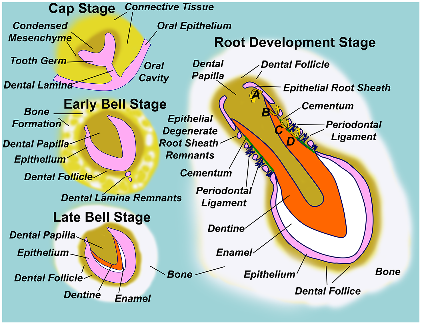
Follow the dental lamina, enamel organ, dental papilla and dental follicle through development. Locate the epithelial root sheath in the root-development panel.
Diagram illustrating normal tooth formation.png · Sarrafpour B, Swain M, Li Q, Zoellner H · CC BY 4.0. None; reproduced without alteration.
Which structure gives rise to dentine and pulp?
The dental papilla. The enamel organ gives rise to enamel; the dental follicle contributes to supporting periodontal tissues.
Pharyngeal arches explain adult nerve patterns
Each pharyngeal arch contains a characteristic cranial nerve, muscular component and skeletal contributions. First-arch muscles include mastication, mylohyoid and anterior digastric and receive V3 motor supply. Second-arch muscles include facial expression, posterior digastric and stylohyoid and receive VII. Stylopharyngeus is associated with the third arch and IX; fourth- and sixth-arch pharyngeal and laryngeal muscle patterns involve branches of X.
The anterior tongue mucosa develops largely from first-arch swellings, explaining lingual V3 general sensation. Posterior tongue mucosal development involves more caudal arches, particularly the third, and is associated with IX. Most tongue muscles arise from migrating occipital myotomes and retain XII motor supply. Development therefore explains why touch, taste and movement need not share one nerve.
Tooth & supporting tissues
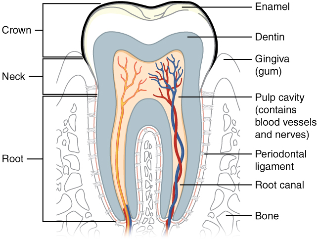
Trace the pulp and root canal to the apical region, then identify the periodontal ligament and supporting bone. Relate these structures to avulsion, apical pathology and extraction. The original uses the spelling “dentin”; cementum is not separately labelled.
Tooth & supporting tissues · OpenStax College · CC BY 3.0. Original image; displayed at a responsive size. Original publication.
Palate formation and cleft consequences
The primary palate develops anterior to the incisive foramen. The secondary palate forms from paired palatal shelves growing from the maxillary prominences. They elevate above the tongue and join each other, the primary palate and nasal septum. This creates separation between oral and nasal cavities, while posterior soft-palate muscle organisation supports speech and swallowing.
A cleft may involve lip, alveolus, hard palate or soft palate in different combinations. Its clinical effects extend beyond a visible gap: feeding, hearing, speech, dental development, eruption and maxillary growth can be affected. Care is therefore coordinated over development. A submucous cleft can preserve surface mucosa despite a deeper structural defect, so appearance alone may not explain function.
Palatal development: historical embryo view
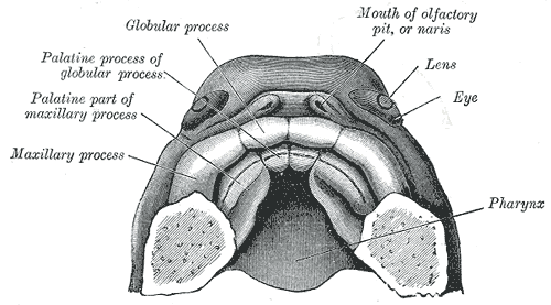
Find the paired palatal processes and the anterior intermaxillary region. The historical labels use “globular process”; interpret these with current descriptions of the medial nasal prominences and primary palate. Use the chapter for modern developmental timing and fusion terminology.
Gray50.png · Henry Vandyke Carter, after His; reproduced in Gray’s Anatomy (1918), plate 50 · Public domain. None; original image reproduced without alteration.
Which structures form the secondary palate?
The paired lateral palatal shelves, arising from the maxillary prominences, elevate and fuse with each other and with the primary palate and nasal septum.
Bud, cap and bell: shape before hard tissue
The dental lamina forms epithelial thickenings at tooth sites. Bud-stage epithelial growth becomes a cap-shaped enamel organ over condensed ectomesenchyme, followed by the bell stage as crown form and cell specialisation develop. The enamel organ, dental papilla and dental follicle are distinct parts of the tooth germ. Reciprocal signals between compartments coordinate their growth rather than one compartment independently manufacturing the whole tooth.
Inner enamel epithelial cells differentiate towards ameloblasts and induce peripheral papilla cells to become odontoblasts. Dentine matrix deposition begins before enamel deposition. The papilla forms dentine and pulp; the follicle contributes cementum, periodontal ligament and alveolar supporting tissues. Disruption of initiation may affect tooth number; disruption of morphodifferentiation may alter tooth size or shape.
| Developing structure | Main contribution | Useful clinical connection |
|---|---|---|
| Enamel organ | Ameloblasts and enamel formation | Defect distribution reflects enamel formation timing |
| Dental papilla | Odontoblasts, dentine and pulp | Dentine and pulp form a linked developmental unit |
| Dental follicle | Supporting periodontal tissues | Do not confuse follicle with pulp-forming papilla |
| Hertwig’s epithelial root sheath | Root pattern and induction of radicular dentine | Crown form precedes root elongation |
Odontomas and other calcified jaw lesions
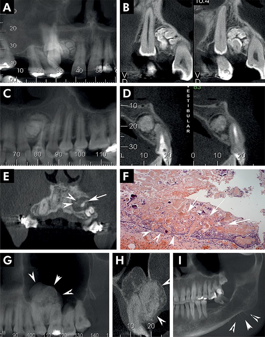
Use the panel key to compare the source diagnoses. The historical label CCOT corresponds here to calcifying odontogenic cyst.
Panel key
- A–B
- Compound odontoma.
- C–D
- Complex odontoma.
- E–F
- Calcifying odontogenic cyst (source label: CCOT).
- G–H
- Adenomatoid odontogenic tumour.
- I
- Source example of osteosarcoma.
Periapical radiopaque-hyperdense jaw lesions 4.jpg · Brunno Santos Freitas Silva, Mike Reis Bueno, Fernanda P. Yamamoto-Silva, Ricardo Santiago Gomez, Ove Andreas Peters and Carlos Estrela · CC BY 4.0. Reproduced without alteration. Labels, arrows and circles, where present, belong to the source image. Original publication.
Does calcification establish benignity?
No. Compare the entire lesion and clinical findings.
Matrix secretion, mineralisation and developmental defects
Ameloblasts secrete an enamel matrix that subsequently matures as mineral content rises and matrix proteins are removed. Hypoplasia describes reduced enamel quantity, such as pits or grooves; hypomineralisation describes defective quality of enamel that may initially have normal thickness. Post-eruptive breakdown can make a qualitative defect appear to be missing enamel, so interpret the original defect and current damage separately.
Distribution helps reconstruct timing and cause. A local injury to a primary predecessor may affect one permanent successor, whereas a systemic disturbance can affect teeth forming at the same time. Inherited disorders can affect broad patterns across a dentition. These are differential clues, not a diagnosis from appearance alone. Ask about family history, childhood illness, trauma and the teeth affected.
Root formation, eruption and primary-tooth resorption
After crown formation, Hertwig’s epithelial root sheath develops from the cervical loop and shapes root development while inducing radicular dentine. Subsequent interaction with follicular cells permits cementum formation. Root elongation, apical maturation and eruption overlap, but eruption is not driven simply by an already completed root pushing upwards.
Eruption requires coordinated remodelling of the eruption pathway and supporting tissues. A permanent tooth commonly emerges before the apex is mature. Primary root resorption permits exfoliation, while an absent or displaced successor can alter that process. Retention, obstruction, ectopia, lack of space and ankylosis are therefore separate explanations for a tooth that is not appearing as expected.
Assess a developmental or eruption discrepancy
Compare the affected tooth with its contralateral partner and expected sequence rather than using age as an absolute deadline. Record retained primary teeth, available space, local swelling or palpable tooth position, previous trauma and relevant family history. Justified imaging can establish presence, developmental stage, orientation and obstruction.
An unerupted tooth may be normally formed but displaced; an erupted tooth may have a formation defect despite an ordinary eruption date. Identify which problem is present before choosing observation, preventive protection, space management or specialist assessment. Document the expected milestone and review interval so that “wait and see” has a defined purpose.
A distinction to remember
Hypoplasia is a quantity defect; hypomineralisation is a quality defect. Eruption failure is another problem and does not by itself show that crown formation was abnormal.
An upper permanent incisor is unerupted while its counterpart is present. The primary predecessor was traumatised in early childhood. What questions should guide investigation?
Reveal the reasoning
Establish whether the permanent tooth is present, its developmental stage and direction, available space and any obstruction or shape disturbance. Childhood trauma increases the relevance of a local developmental or eruption disturbance, but does not establish which mechanism is responsible. Compare clinical palpation with justified imaging.
Guidance to read alongside this chapter
Test your recall
Which tissues form enamel, dentine and pulp?
Show answer
Ameloblasts from the enamel organ form enamel. Odontoblasts from the dental papilla form dentine, and the remaining papilla contributes the pulp. The dental follicle contributes periodontal supporting tissues, so it should not be substituted for the papilla in this sequence.