
On-call referrals and triage
Make an immediate safety decision, then answer the referral question.
What to expect
Calls may come from ED, wards, dentists, GPs and other hospitals. Some concern a routine facial laceration; others involve a threatened airway or major trauma. Take the patient’s location and a callback number first. Establish whether they are stable, whether a senior is already present and whether immediate emergency help is needed.
What to know
Use an ABCDE approach and current observations rather than relying on a diagnosis label. Ask about breathing difficulty, voice change, drooling, swallowing, trismus, swelling trajectory, bleeding, eye symptoms, neurological change, anticoagulants and comorbidity. For trauma, clarify mechanism, time, loss of consciousness, cervical-spine concern, occlusion, visual symptoms and imaging already obtained.
A practical approach
State the immediate action, urgency and destination. A concise handover includes age, problem, current observations, red flags, examination, relevant imaging, treatment already given and the question for OMFS. Record advice and who accepted responsibility. Reassess when the patient arrives: a telephone description can be incomplete or outdated.
Safety point
A normal temperature or small visible swelling does not exclude deep-space infection. Escalate airway features, rapidly progressive swelling, uncontrolled haemorrhage, visual compromise or physiological deterioration immediately.
Structured assessment and differential diagnosis
A strong assessment produces a clinical question, a ranked differential and a safe next step. Collect discriminating information rather than an unstructured list of positive findings.
History that changes the plan
Characterise onset, duration, progression, site, radiation, triggers, relieving factors and functional effects. Pain related to biting, temperature, meals or light touch points towards different mechanisms. Ask about previous episodes and treatment response, but do not let a prior label replace reassessment. Establish the patient’s concerns and desired outcome.
Record medicines with dose and indication, over-the-counter products, allergy reaction and timing, bleeding history, medical stability, previous anaesthetic problems and relevant social factors. Smoking, alcohol, nutrition, support at home and ability to attend follow-up can affect both risk and the practical treatment choice. Ask directly about swallowing, breathing, voice change and rapidly progressive swelling when infection is possible.
Trigeminal nerve & dental branches
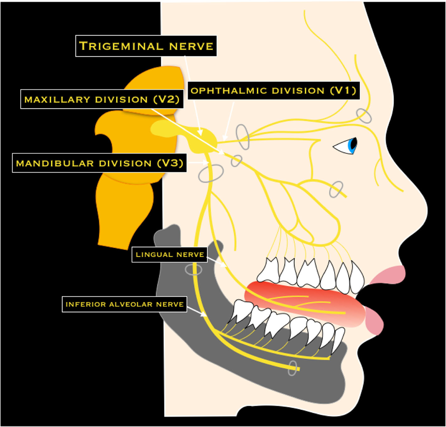
Trace V1, V2 and V3, then the lingual and inferior alveolar nerves. Connect each to the sensory examination after lower third-molar surgery. This is a branch overview, rather than an injection-placement diagram.
Trigeminal nerve & dental branches · Nicola Romano, Margherita Federici and Antonio Castaldi · CC BY 4.0. Original image; displayed at a responsive size. Original publication.
Examine systematically
| Domain | Observe / test | Why it matters |
|---|---|---|
| Extraoral | Symmetry, swelling, skin change, nodes and gland findings | Extent, malignancy clues and non-dental causes. |
| Neurological | Relevant cranial nerve sensation and movement | A baseline for consent and meaningful postoperative comparison. |
| Function | Opening, excursions, occlusion, speech and swallowing | Deep infection, trauma, joint restriction and airway implications. |
| Intraoral | Mucosa, floor of mouth, tongue, duct openings, periodontal and dental findings | Correlate the symptom with a source; inspect the whole mouth. |
| Tooth tests | Percussion, palpation, mobility, probing and appropriate sensibility tests | Differentiate pulpal, periodontal, periapical and structural causes. |
Head and neck lymphatic glands (Gray's Anatomy, plate 602)
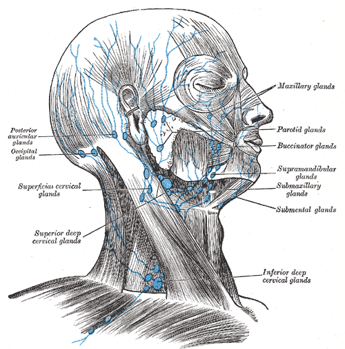
Find the submental and submandibular groups and follow drainage toward the cervical nodes. In this historical figure, ‘glands’ means lymph nodes and ‘submaxillary’ means submandibular. Use current cancer guidance for modern oncological neck levels.
Head and neck lymphatic glands (Gray's Anatomy, plate 602) · Henry Vandyke Carter; reproduced in Henry Gray, Anatomy of the Human Body (1918), plate 602 · Public domain. Unchanged published image; displayed responsively.
Rank and safety-check the differential
Separate the most likely diagnosis from the most serious plausible alternative. For example, a non-healing socket may reflect a local healing problem, but malignancy, necrosis or an underlying bone lesion may require exclusion. Use duration, risk factors, examination and imaging to discriminate; “infection” is not an adequate endpoint if the source is unclear.
Document pertinent negative findings, the working diagnosis and what would trigger reassessment. Unexplained paraesthesia, persistent induration, suspicious nodes, progressive trismus and airway symptoms should change urgency. If a finding cannot be explained by the proposed diagnosis, investigate it rather than forcing it into the original story.
Move from information to a clinical decision
Likely cause
- Symptom pattern and examination
- Find a coherent source
- Plan a test or definitive treatment
Serious alternative
- Identify red flags
- Ask what must not be missed
- Escalate or investigate promptly
Uncertainty
- Identify the unexplained finding
- Choose a discriminating investigation
- Review with an explicit safety net
A distinction to remember
A positive finding is useful only in context. One tender tooth does not explain an otherwise unexplained neck lump or sensory deficit.
A patient has lower lip paraesthesia and a poorly defined mandibular lesion. Why is “dental infection” an incomplete assessment?
Reveal the reasoning
The sensory deficit and destructive appearance raise serious alternatives. Establish vitality and the clinical source, describe the lesion and arrange appropriate urgent specialist investigation rather than treating repeatedly with antibiotics.
Sedation events, LA toxicity and emergency handover
Procedure-related deterioration needs the same physiological priorities plus a precise medicine history. The formulation and timing can be essential to the receiving team.
Distinguish plausible causes
| Problem | Clues | Response priority |
|---|---|---|
| Oversedation / respiratory depression | Reduced effort, contact or airway tone after depressant medicines | Stop administration, call help and support airway / ventilation. |
| LA systemic toxicity | Neurological symptoms, seizures or cardiovascular change after LA exposure | Stop LA and use the current specialist LAST pathway. |
| Vasovagal episode | Trigger, pallor, sweating and compatible recovery pattern | Assess and position; revisit diagnosis if recovery is atypical. |
| Adrenaline-related symptoms | Temporal relationship to injection with palpitations / tremor | Assess the patient; do not dismiss serious cardiovascular findings. |
| Anaphylaxis | Airway / breathing / circulation compromise after a plausible exposure | Emergency anaphylaxis treatment; rash is not required. |
Do the supportive steps immediately
Stop treatment and the implicated medicine, summon suitable support and reassess ABCDE. For respiratory compromise, airway and ventilation support are immediate priorities; reversal medicines are adjuncts. Continued observation is needed after reversal because the original sedative or opioid can outlast its antagonist.
For suspected LAST, follow the current lipid-emulsion and modified-resuscitation checklist with trained anaesthetic / resuscitation help. The protocol includes important differences from generic arrest medicine use. A printed current emergency protocol and trained team are more useful than an isolated memorised lipid quantity.
Handover and incident learning
Use a structured handover: situation, background, assessment and required action. Include the event time, procedure, exact medicine / concentration / route / total dose, other exposures, observations, interventions and response. Report what is uncertain, including possible retained or aspirated material, rather than omitting it.
After stabilisation, document openly, support the patient and arrange the appropriate review and reporting. Debrief the team, identify practical system changes and rehearse emergency roles. A completed incident form does not itself improve readiness unless the learning changes practice.
A useful handover preserves the critical facts
Situation
- What happened and when
- Current ABC findings
- Urgent support required
Exposure
- Exact medicine and formulation
- Route, dose and timing
- Other medicines / possible causes
Response
- Interventions already given
- Physiological response / unresolved issues
- Transfer, monitoring and follow-up plan
A distinction to remember
“Three cartridges” is an incomplete dose handover: volume and concentration vary. Provide the actual formulation and calculated total amount.
What dose information should accompany suspected LA toxicity?
Reveal the reasoning
The exact local anaesthetic, concentration, cartridge / injected volume, total mass, vasoconstrictor formulation, route and time, plus other doses and the clinical course.
Guidance to read alongside this chapter
Test your recall
Which details come first in an on-call phone referral?
Show answer
The patient’s location, callback number, current stability and whether immediate senior or emergency help is required.